Equipment Management - LABS FOR LIFE PROJECT - INDIA
Histopathology and cytology equipment, focusing on the tissue processor, important parts, maintenance, and steps in tissue processing. Understand the significance of these tools in the microscopic examination of tissues for disease manifestations.
Download Presentation

Please find below an Image/Link to download the presentation.
The content on the website is provided AS IS for your information and personal use only. It may not be sold, licensed, or shared on other websites without obtaining consent from the author. Download presentation by click this link. If you encounter any issues during the download, it is possible that the publisher has removed the file from their server.
E N D
Presentation Transcript
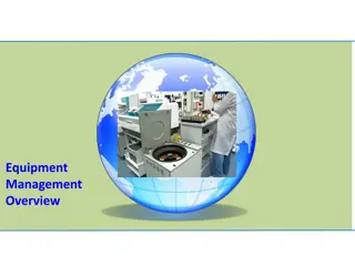
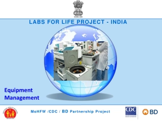
LABS FOR LIFE PROJECT - INDIA Equipment Management MoHFW /CDC / BD Partnership Project
HISTOPATHOLOGY AND CYTOLOGY EQUIPMENT Histopathology Compound of three Greek words: histos "tissue", pathos "suffering", and logia "study of It refers to the microscopic examination of tissue in order to study the manifestations of disease. In clinical medicine, histopathology refers to the examination of a biopsy or surgical specimen by a pathologist, after the specimen has been processed and histological sections have been placed onto the glass slide. Histopathological examination of tissues starts with surgery, biopsy, or autopsy. The tissue is removed from the body and then placed in a fixative which stabilizes the tissues to prevent decay. The most common fixative is formalin (10% formaldehyde in water).
HISTOPATHOLOGY AND CYTOLOGY EQUIPMENT TISSUE PROCESSOR A tissue processor is used to prepare tissue samples for analysis by fixing, staining, dehydrating or decalcifying them. These instruments allow the specimens to be infiltrated with a sequence of different solvents finishing in molten paraffin wax. The specimens are in an aqueous environment to start with (water-based) and must be passed through multiple changes of dehydrating and clearing solvents (typically ethanol and xylene) before they can be placed in molten wax (which is hydrophobic and immiscible with water). The duration and step details of the processing schedule chosen for a particular batch of specimens will depend on the nature and size of the specimens. Schedules can be as short as one hour for small specimens or as long as twelve hours or more for large specimens. Types of processors: 1. The tissue-transfer (or dip and dunk ) equipment where specimens are transferred from container to container to be processed 2. The fluid-transfer (or enclosed ) types where specimens are held in a single process chamber or retort and fluids are pumped in and out as required.
HISTOPATHOLOGY AND CYTOLOGY EQUIPMENT Important Parts and Maintenance: (a) Tissue containers - These are also the cassettes. The tissue to be processed is placed in an appropriate container, together with a label and the lid snapped on. (b) Beakers and wax baths - Most equipment are equipped with ten beakers and 2 wax baths thermostatically controlled. (c) Stirring mechanism - The basket is attached to the arms of the equipment on which one arm is designed in such a manner so as to bring about the rotation of the basket nearly at the rate of one revolution per minute. (d) Timing mechanism - Timer is meant to keep the tissue in different reagents and wax for an optimum time. (e) Persons responsible for operating the instrument should be properly trained in its use and training record to be documented. (f) Instrument operating manual should be as per the manufacturer s instructions and SOP and calibration procedure should be done as defined by them. It would vary from instrument to instrument as per the specification. Automated Tissue Processor
HISTOPATHOLOGY AND CYTOLOGY EQUIPMENT Some important steps of tissue processing are as below Dehydration Melted paraffin wax is hydrophobic (immiscible with water), most of the water in a specimen must be removed before it can be infiltrated with wax. This process is commonly carried out by immersing specimens in a series of ethanol (alcohol) solutions of increasing concentration until pure, water-free alcohol is reached. Ethanol is miscible with water in all proportions so that the water in the specimen is progressively replaced by the alcohol. A series of increasing concentrations is used to avoid excessive distortion of the tissue. Use of copper sulphate in final alcohol: A layer of anhydrous CuSO4 is placed at the bottom of a dehydrating bottle or beaker and is covered with 2-3 filter papers of approximate size to prevent staining of the tissue. Anhydrous CuSO4 removes water from alcohol as it in turn removes it from tissues. Anhydrous CuSO4 is white in color while the hydrated form is blue.
HISTOPATHOLOGY AND CYTOLOGY EQUIPMENT Clearing The term clearing was chosen because many (but not all) clearing agents impart an optical clarity or transparency to the tissue due to their relatively high refractive index. We now have to use an intermediate solvent that is fully miscible with both ethanol and paraffin wax. This solvent will displace the ethanol in the tissue, then this in turn will be displaced by molten paraffin wax. This stage in the process is called clearing and the reagent used is called a clearing agent . Another important role of the clearing agent is to remove a substantial amount of fat from the tissue which otherwise presents a barrier to wax infiltration. A popular clearing agent is xylene and multiple changes are required to completely displace ethanol. A typical clearing sequence for specimens not more than 4mm thick would be: Xylene 20 min Xylene 20 min Xylene 45 min
HISTOPATHOLOGY AND CYTOLOGY EQUIPMENT Wax infiltration The tissue can now be infiltrated with a suitable histological wax. Paraffin wax-based histological waxes are the most popular. A typical wax is liquid at 60 C and can be infiltrated into tissue at this temperature then allowed to cool to 20 C where it solidifies to a consistency that allows sections to be consistently cut. These waxes are mixtures of purified paraffin wax and various additives that may include resins such as styrene or polyethylene. It should be appreciated that these wax formulations have very particular physical properties which allow tissues infiltrated with the wax to be sectioned at a thickness down to at least 2 m, to form ribbons as the sections are cut on the microtome, and to retain sufficient elasticity to flatten fully during flotation on a warm water bath. A typical infiltration sequence for specimens not more than 4mm thick would be: Wax 30 min Wax 30 min Wax 45 min
HISTOPATHOLOGY AND CYTOLOGY EQUIPMENT Maintenance Use of an inappropriate processing schedule or the making of a fundamental mistake (perhaps in replenishing or sequencing of processing reagents) can result in the production of tissue specimens that cannot be sectioned and therefore will not provide any useful microscopic information. Although mechanical or electrical faults occasionally occur in tissue processors, processing mishaps where tissues are actually compromised, mainly occur because of human error. The need for UPS is very vital in ensuring uninterrupted carousal movements. Interruption to carousal movement may result in the basket hanging above and drying out and tissue damage. Similarly, prolonged immersion in Xylene, Isopropyl alcohol etc. will result in hardening of tissue. It is important to have a programming worksheet for the reagent change schedule for reference. The need for change depends on the volume of work. The number of blocks processed before change of the alcohols, xylenes, wax etc. should be considered while a procedure for this is developed and documented
HISTOPATHOLOGY AND CYTOLOGY EQUIPMENT Maintenance Since most tissue processors are run during night hours, there should be arrangement for interventions in case of breakdown. Interruption to carousal movement may result in the basket hanging above and drying out and tissue damage. Pronged immersion in Xylene, Isopropyl alcohol etc. will result in hardening of tissue. It is important to have a programming worksheet for the reagent change schedule for reference. Mop up spilled reagents immediately. Clean the instrument on a daily basis. Once a month, lift the carousel cover to its upper end position, clean the carousel axle with a cleaning cloth and subsequently apply a thin coat of equipment oil. Have a preventive maintenance done once a year by a service manager authorized. Once the warranty period expires, it is recommended to purchase a service contract. As per NABL 112 Depending on the workload the laboratory must develop a procedure to change the tissue processing fluids and maintain a record of it. A log recording of the time setting schedule for an automatic tissue processor must be maintained. Temperature of the wax bath must be checked and recorded daily.
HISTOPATHOLOGY AND CYTOLOGY EQUIPMENT Monitoring Points to noted Fluid and wax beakers must be filled upto appropriate mark and located in their correct position in the machine. Any spillage of the fluid should be wiped away. Accumulations of wax must be removed from beaker, covers, lids and surrounding areas. Wax bath thermostats should be set at satisfactory levels usually 2-3 C above the melting point of wax. Particular attention should be paid to fastening the processing baskets on the crousel type of machines, if the baskets are shed they will remain in one particular regent for a long period till it gets noticed. Timing should be set with utmost care when loading the machine. Paraffin wax baths should be checked to ensure that the wax is molten Formats to be prepared and the tissue processing data for each cycle should be documented.
HISTOPATHOLOGY AND CYTOLOGY EQUIPMENT MICROTOME: ROTARY MICROTOME A microtome is a tool with blades used to cut extremely thin slices of tissues, known as sections. Several types of microtomes are available - rotary, rocking, base-sledge etc. Microtomes use different kinds of blades depending upon the specimen being sliced and the desired thickness of the sections being cut. The tissue is cut in the microtome at thicknesses varying from 2 to 50 m. The consistently thin samples obtained by microtomy, together with the selective staining of important cell components or molecules allow for the visualization of microscope details in tissues for histopathological analysis. This device operates with a staged rotary action such that the actual cutting is part of the rotary motion. The flywheel in many microtomes can be operated by hand.
HISTOPATHOLOGY AND CYTOLOGY EQUIPMENT Components Microtome base plate or stage Knife holder base Knife holder Cassette clamp or block holder Coarse hand wheel Micron adjustment
HISTOPATHOLOGY AND CYTOLOGY EQUIPMENT Microtome knives Types of microtome knives: Microtome knives are classified by the manner in which they are ground and seen in their cross section. Plane wedge: It is used for paraffin and frozen sections. Plano concave: used for celloid in section since the blade is thin it will vibrate when for used for other harder materials. Biconcave: It is recommended for paraffin section cutting on rocking and sledge type of microtome. Tool edge: This is used with a heavy microtome for cutting very hard tissues like undecalcified bone.
HISTOPATHOLOGY AND CYTOLOGY EQUIPMENT Cleaning and daily maintenance : Before cleaning: Lock the rotary hand wheel. Remove the microtome blade from the knife holder. Remove knife holder base and knife holder for cleaning Cleaning: Remove all sectioning waste with a dry brush. To remove paraffin residue, use paraffin removing product. Clean the outside surfaces with warm soapy water, and dry with a moist cloth. Safety precautions Do not handle the blade with unprotected hands. Microtome with non-disposable knife should have a safety shield Make sure to use forceps and wear gloves. Do not leave the blade exposed during sample loading and unloading. Remember to cover the blade. Cover the blade by using the provided finger guards or a protective foam guard over the blade during mounting/removal of sample. Use forceps to remove the blade and wear two layers of nitrile gloves to help protect your skin Do not skip Personal Protective Equipment (PPE) gloves can minimize or possibly prevent cuts and lacerations
HISTOPATHOLOGY AND CYTOLOGY EQUIPMENT Microtome Knife Sharpening This is done by manual or automatic methods. Two steps: -honing and stropping are employed for knife sharpening Honing: Microtome knives are sharpened against a special stone called Hone . Honing refers to grinding the cutting edge of the knife on a hard abrasive surface to sharpen the knife. Stropping: This is a process of polishing an already fairly sharp edge that may be flexible (hanging) or rigid. . Knife Sharpening Equipment Despite high cost these equipment are popular because they are less time consuming.
HISTOPATHOLOGY AND CYTOLOGY EQUIPMENT Daily: 1.Lock the microtome hand wheel. 2.Remove all section debris, using either alcohol-soaked paper towel or a suitable vacuum system. 3.Carefully remove all specimen trimmings. 4.Remove all specimens. 5.Remove all blades or knives. 6.Remove all used specimen holders and soak in warm soapy water to ensure that all cryocompound is removed; air or oven dry. Weekly: 1.Empty fluid container (this fluid is collected during the daily defrost cycles). 2.Thoroughly clean and lubricate all contact surfaces of the knife holders. Monthly: 1.Spindle lubrication: ensure that the hand-wheel of the microtome is locked, using the coarse advance bring the microtome spindle (object head) forward. 2.Using a disposable pipette place a few drops of low-temperature oil (supplied with the cryostat) onto the rear (close to the microtome housing) upper surface of the spindle 3.Retract the spindle to its home position. 4.Check the side compressor vents to remove all dust etc. Annual: Have the instrument serviced by Manufacturer trained and approved service engineers.
HISTOPATHOLOGY AND CYTOLOGY EQUIPMENT TISSUE FLOATATION BATH The tissue floatation/ water baths are used to float paraffin ribbons, to stretch sections and remove wrinkles and folds before placing sections on the slide. Simply sticking paraffin ribbons on slides does not work. A warm water bath allows tissue to relax and smooth out prior to being mounted on a glass slide. The warmth also makes the paraffin stick to the glass slides. These water baths are filled with distilled water, heated to a temperature 5-10 C below the melting point of paraffin, and the water bath is usually kept at 40-50 C. This is an optimal temperature range for various types of paraffins. Hard paraffin will require a higher water temperature to relax, while softer paraffins will benefit from a lower water temperature since they may disintegrate at higher temperatures.
HISTOPATHOLOGY AND CYTOLOGY EQUIPMENT Maintenance Water baths are equipment whose maintenance is simple. The recommended routines mainly focus on the cleaning of external components. 1.Water must be kept clean and changed at least daily. 2.Keep empty reservoir covered to prevent fibers and dust from settling and becoming a potential contaminant. 3.Water surface must be skimmed with a laboratory-grade wipe to prevent contamination of debris between each ribbon. 4.Temperature of the bath should be checked and recorded daily. 5.Check the thermometer or temperature controls every three months using known standards. If no reference standard is available, use an ice/water mixture and/or boiling water. Note that the thermometer or the water bath temperature controls should also be checked when the equipment is first installed after purchase. Limitations The water bath may introduce artifacts to sections. As per NABL 112- 1.The fluid in the flotation bath shall be changed at least once a day. 2.The surface of the water bath shall be skimmed regularly during section cutting to remove floaters.
HISTOPATHOLOGY AND CYTOLOGY EQUIPMENT CRYOSTATS The tissue can be rapidly frozen and kept frozen while sections are cut using a cryostat microtome (a microtome in a freezing chamber). These are called frozen sections . Frozen sections can be prepared very quickly and are therefore used when an intra-operative diagnosis is required to guide a surgical procedure or where any type of interference with the chemical makeup of the cells is to be avoided. Types Several types of cryostats are commercially available and can be categorized as follows: a. Single compressor (chamber cooling only) b. Double compressor (chamber and object cooling) c. Manual sectioning d. Motorized sectioning The normal working chamber temperature is from 0 C to 35 C, the limiting factor being the type of compressor and refrigerant used. Cryosectioning at temperatures lower than 35 C requires the use of a cryogen such as liquid nitrogen.
HISTOPATHOLOGY AND CYTOLOGY EQUIPMENT Maintenance Cryostats must be located in a draught-free, humidity-controlled area, with clearance of 30 cm, on all sides to ensure that there is unrestricted air movement around the instrument. All accessories; knife holder, antiroll guide, brush trays, etc. should be placed into the chamber prior to the instrument being turned on. Always clean the equipment between users and at the end of a session. Perform a comprehensive decontamination by removing the stage and allowing it to come to room temperature before cleaning. Allow it to dry completely before returning it to the cryostat. Use a pair of forceps to carefully remove the knife. Either discard the knife directly in a sharps container or if re-using it, place in a container of disinfectant to soak. Specimen shavings should be removed after each use. The waste tray that is underneath the microtome spindle should be carefully lifted out and the shavings/debris placed into a biohazard bag. Other shavings and specimen debris can be wiped up using either paper towel or a gauze swab soaked in 70% alcohol. During this procedure it is important to minimize aerosols and to prevent sectioning debris from spilling onto open surfaces. Use a cloth, manipulated with forceps where possible, to scrub and decontaminate. If needed, use a handled brush to access hard to reach areas, though a brush is more likely to splatter so it is necessary to use facial protection. Soak up residual disinfectant and rinse/dry with 95% ethanol. Let air dry.
HISTOPATHOLOGY AND CYTOLOGY EQUIPMENT Safety precautions 1.Histology specimen preparation in the BSL-1 and BSL-2 Lab involves very sharp blades and hence strict adherence to safety measures is critical. 2.Do not handle the blade with unprotected hands. 3.Make sure to use forceps and wear gloves. Do not leave the blade exposed during sample loading and unloading. Remember to cover the blade. 4.Cover the blade by using the provided finger guards or a protective foam guard over the blade during mounting/removal of sample. 5.Use forceps to remove the blade and wear two layers of nitrile gloves to help protect your skin 6.Do not skip Personal Protective Equipment (PPE) gloves can minimize or possibly prevent cuts and lacerations. As per NABL 112 Frozen section/squash smear: a.A specific area should be demarcated for performing frozen sections. b.Fresh tissue received for frozen section should be treated as infective and universal precautions should be taken. c.Frozen sections/squash smears should be recorded like other specimens in the request form. Left over tissue must be processed for permanent section. d.The turnaround time for frozen section/squash smears should not exceed 30 minutes. e.Frozen section/squash smears shall be retained and filed along with the permanent sections for the stipulated time.
HISTOPATHOLOGY AND CYTOLOGY EQUIPMENT Prion disease suspected specimens: In a suspected case of prion disease, facilities should be available for safe handling of specimens. The biopsy specimen shall be considered as bio-hazardous and transferred to concentrated formic acid (96%) for 48 hours, subsequently to 10% formalin for 24 hours and then processed. The blocks should be labeled bio hazardous. The trimmings of the block shall be disposed by incineration. All instruments used for sectioning should be left in 2M NaOH for 1 hour and washed in running water for 15 minutes and reused. The microtome should be wiped clean with 2M NaOH and left for 1 hour. Subsequently the instrument should be wiped clean with tap water followed by alcohol before reuse.
HISTOPATHOLOGY AND CYTOLOGY EQUIPMENT ANTIGEN RETRIEVAL SYSTEMS In the majority of cases, tissue specimens for immune histochemical (IHC) staining are routinely fixed in formalin and subsequently embedded in paraffin. Suboptimal methods of antigen retrieval and buffer used can result in poor antigen retrieval or over expression or overcalling Several methods of antigen retrieval are available 1. 2. 3. 4. 5. 6. 7. Water Bath Methods Pressure Cooker Heating Autoclave Heating Microwave Oven Heating Proteolytic Pre-treatment Combined Proteolytic Pre-treatment and Antigen Retrieval Combined Deparaffinization and Antigen Retrieval
HISTOPATHOLOGY AND CYTOLOGY EQUIPMENT CYTOCENTRIFUGATION Cytocentrifugation is centrifugation using a Cytocentrifuge. Using centrifugal principles, the cells are deposited onto a clearly-defined area of a glass slide. The monolayer distribution enhances the morphological appearance of the cells present. It is suitable for complete range of body fluids like CSF, Pleural fluid, Sputum, Urine, Synovial fluids, Peritoneal fluids, Bronchial washings, Fine needle aspirates etc. The technique is a quick method to collect and concentrate above mentioned fluids that contain a low number of target cells. It provides more cells than which would be present in a wedge smear preparation on the same sample and therefore, provides for greater precision in counting. A prominent advantage of the Cytocentrifuge is the much smaller area to review and consequently the shortened time required to read the slides. Cytocentrifugation also constructively flattens cells for excellent nuclear presentation.
HISTOPATHOLOGY AND CYTOLOGY EQUIPMENT Principle The principle of all cytocentrifugation techniques is that the cell is denser than the suspending fluid and under an applied force the cell will have greater momentum than the fluid. It produces good cell capture and representation of all cell types present in homogeneous liquid samples. Key components 1. 2. 3. 4. 5. 6. 7. 8. Power ON-OFF Switch ON-OFF Switch Lamp Lid Lid Latch Rotor Bowl Rotor Lid Seal Control Panel .
HISTOPATHOLOGY AND CYTOLOGY EQUIPMENT Maintenance Daily 1.Wipe the external surfaces with a clean cloth moistened with neutral detergent. Pat dry any excessively moist areas. 2.Wipe the rotor bowl with a cloth wet with disinfecting solution. Then clean the rotor bowl by wiping it with a clean cloth moistened with neutral detergent. 3.Disconnect the drain hose and place a pan under the drain port of the instrument. Thoroughly rinse the rotor bowl with clear water. Completely dry the rotor bowl with a clean, lint-free cloth. 4.Do not use excessive quantities of liquid when cleaning the rotor bowl. 5.All the reusable components like specimen chamber holder, rubber gasket, specimen chamber, and specimen chamber cap should be carefully decontaminated and cleaned prior to use. All components should be soaked in an approved disinfectant. After soaking the components in an approved disinfectant, they should be cleaned in hot water and detergent. The water temperature should not exceed 40 C. As per NABL 112 1.All equipment such as centrifuges capable of creating bio-hazardous aerosols should be used in extractor cabinets or rooms fitted with extractor facilities. 2.The laboratory performing Cytopathology tests on CSF must use Cytocentrifuge for processing the sample.
HISTOPATHOLOGY AND CYTOLOGY EQUIPMENT LIQUID BASED CYTOLOGY Liquid-based cytology is a method of preparing samples for examination in cytopathology. The sample is collected, normally by a small brush, in the same way as for a conventional smear test, but rather than the smear being transferred directly to a microscope slide, the sample is deposited into a small bottle of preservative liquid. At the laboratory, the liquid is treated to remove other elements such as mucus before a layer of cells is placed on a slide. The technique allows more accurate results. It has gained popularity as a method of collecting and processing both gynecologic and non-gynecologic cytological specimens. It achieves a diagnostic sensitivity as accurate as conventional preparations especially for its excellent cell preservation and for the lack of background which decrease the amount of inadequate diagnoses.
DIAGNOSTIC TECHNIQUES Conventional Pap Colposcopy HPV Molecular Test Others : VIA / VILI
CONVENTIONAL PAP Sample Collection Specimen Smearing & Slide Fixing Standardization Discard Collection Brush ( important diagnostic material) Slide Staining ( in Lab) Microscopic visualization of the larger area on the slide. Conventional Pap
HISTOPATHOLOGY AND CYTOLOGY EQUIPMENT Conventional Pap smears can have false-negative and false-positive results because of inadequate sampling and slide preparation, and errors in laboratory detection an interpretation. LBC rinses Cervical cells in preservatives so that blood and other potentially obscuring material can be separated. It also allows for additional testing of the sample, such as for human papillomavirus (HPV).
