Overview of Superficial Fungal Infections of the Skin
Superficial fungal infections of the skin are categorized into superficial, cutaneous, and subcutaneous types. Superficial mycoses affect the stratum corneum and include conditions like tinea versicolor, tinea nigra, and piedra. Tinea versicolor is a chronic infection caused by Malassezia furfur, while tinea nigra presents as painless brown or black patches. Piedra is an asymptomatic infection of the hair shaft. Diagnosis involves skin scrapings and cultural identification of the causative fungi.

Overview of Superficial Fungal Infections of the Skin
PowerPoint presentation about 'Overview of Superficial Fungal Infections of the Skin'. This presentation describes the topic on Superficial fungal infections of the skin are categorized into superficial, cutaneous, and subcutaneous types. Superficial mycoses affect the stratum corneum and include conditions like tinea versicolor, tinea nigra, and piedra. Tinea versicolor is a chronic infection caused by Malassezia furfur, while tinea nigra presents as painless brown or black patches. Piedra is an asymptomatic infection of the hair shaft. Diagnosis involves skin scrapings and cultural identification of the causative fungi.. Download this presentation absolutely free.
Presentation Transcript
Lecture Title: Fungal Infections of the skin Superficial and cutaneous infections ( Microbiology) Lecturer name: Dr. Ahmed M. Albarrag Lecture Date: Dec-2011
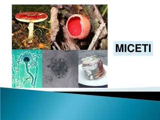
Skin fungal infections Clinical Skin fungal infections are generally divided into : (1) Superficial, including tinea versicolor, tinea nigra and piedra (2) Cutaneous, including Dermatophytosis, Candidiasis of skin ,mucosa, and nails and others (3) Subcutaneous, including mycetoma, sporotrichosis, chromoblastomycosis; and others
Superficial Mycoses Defined as infections in which a fungal pathogen is restricted to the stratum corneum, with little or no tissue reaction. These affect the uppermost dead layers of skin or hair shaft. They are painless and usually do not provoke the immune system They include: 1- Tinea versicolor 2- Tinea nigra 3- Piedra
Superficial Mycoses Tinea Versicolor Tinea versicolor is a long-term (chronic) fungal infection of the skin Patches of brown or discolored skin with sharp borders and fine scales. The patches are often dark reddish-tan in color The most common sites: The back, underarms, upper arms, chest, and neck. Affected areas do not darken in the sun there may be hypopigmentation or hyperpigmentation of skin. Usually asymptomatic Etiology: Malassezia furfur It is a Yeast, Lipophilic Normal flora of skin
Tinea Versicolor Diagnosis: Skin scraping Potassium hydroxide (KOH) Positive for short hyphae and spores (Spaghetti and meatballs) Culture: Malassezia furfur It is a Yeast, Lipophilic To grow, oil should be added to the media
Superficial Mycoses Tinea nigra Painless macules or patches with brown or black color Usually located on palm of hand or sole of foot. Acquired by Piercing of skin with plant material in Agricultural soil. Etiology: Exophiala werneckii Dematiaceous filamentous fungus Laboratory Diagnosis: Skin scrapings: In 10% or 20% KOH will show brown septate hyphae Culture on SDA & Mycobiotic: growth of dematiaceous fungus. Identify my microscopic appearance of conidia
Superficial Mycoses Piedra Asymptomatic infection of the hair shaft, Nodules on hair shaft On scalp hair / mustache, beard Black piedra Dark pigmented nodules. Hard and firmly attached to hair shaft, Eiology : Piedraia hortae White piedra Lightly pigmented, white to brown nodules, Soft, loosely attached Etiology:Trichosporon beigelii yeast Pseudohyphae, arthrospores Lab Diagnosis: Hair with nodule Direct microscopy: 10% -20% KOH Culture : on Mycobiotic & SDA
Treatment of Superficial infections 2% salicylic acid, 3% sulfur ointments, whitfield s ointment Ketoconazole Piedra: Cutting or shaving the hair Or apply 2% salicylic acid Or 3% sulfur ointment. Nizoral shampoo (contains Ketoconazole) Antifungal agents Topical Systemic
Dermatophytoses Fungal infections of the Keratinized tissues of the body Scalp, glabrous skin, and nails caused by a closely related group of fungi known as dermatophytes . They are primary pathogens Contagious transmitted through infected scales hyphae or arthroconidia on the skin. direct contact between infected humans or animals (goats, sheep, camel, cows, horses Transfer form on area to the body to another, Familial cross infection occurs Tinea or Ringworm T.capitis T.corporis : T.pedis scalp glabrous skin foot (Athlete s foot) T.cruris: T. unguium nail T.barbae beard T.manuum hand groin
Etiology Dermatophytes A group of related fungi called dermatophytes (filamentous fungi) Primary pathogens Microsporum - infections on skin and hair Epidermophyton - infections on skin and nails Trichophyton - infections on skin, hair, and nails. Geophilic species - keratin-utilizing soil saprophytes (e.g., M. gypseum, T. ajelloi). Zoophilic species - keratin-utilizing on hosts - living animals (e.g., M. canis, T. verrucosum). Anthropophilic species - keratin-utilizing on hosts - humans (e.g., M. audounii, T. tonsurans)
Tinea Capitis Presentations of Tinea Capitis 1. Non-inflammatory 2. Pustular 3. Inflammatory Kerion Favus (=t.favosa) with scutulum (yellow crusts) Using the Wood s lamp on infected hair fluoresce especially microsporum spp. lesions.
Tinea Capitis Diagnosis History Contact with infected person, pets, duration Clinical presentation Broken hairs, black dots, localized, inflammatory, etc. Woods Lamp Blue green. Hair Shaft Exam 10-20% KOH, Endo/Exothrix Culture
Other Identification Tests: 1) Endothrix & Ectothrix hair infection 2) Hair perforation test 3) Urease test 4) Pigment production 5) Nutrient requirement such as Trichophyton series Agar 1-7
Tinea Capitis Treatment Must treat hair follicle Topical , but might be not effective Systemic agents Griseofulvin for children liquid with good taste. Terbinafine. Treat until no visual evidence, culture (-) plus 2 weeks Average of 6-12 weeks of treatment. Examine / treat family in recurrent cases.
General Morphology Onychomycosis General Appearance: Typically begins at distal nail corner Thickening and opacification of the nail plate Nail bed hyperkeratosis Onycholysis Discoloration: white, yellow, brown
Candidaisis of nail Paronychia
Diagnostic Tests KOH Preparations Skin Nails Thin clipping, shaving or scraping Let dissolve in KOH for 6-24 hours. Can be difficult to visualize. Culture often required. Hair Apply KOH Look for fungal elements
Diagnostic Tests KOH Preparations Skin Two slides or slide and #15 blade. Scrape border of lesion. Apply 1-2 drops of KOH and heat gently Examine at 10x and 40x Focus back and forth through depth of field. Look for hyphae Clear, Green Cross cell interfaces Branch, constant diameter. Chlorazol black, Parkers ink can help.
Diagnostic Tests Fungal Cultures Sabouraud dextrose Agar (SDA) DTM (Dermatophyte Test Medium) Yellow to red is (+).
Diagnostic Test: Fungal Culture DTM A special medium for the identification of dematophytes It has pH 5.6, Antibacterial, Antifungal, and Phenol red (Amphoteric dye) Not recommended for use in clinics. Positive Negative Growth and change in color to red
Other Identification Tests: 1) Endothrix & Ectothrix hair infection 2) Hair perforation test 3) Urease test 4) Pigment production in PDA & CMA media 5) Nutrient requirement such as Trichophyton series Agar 1-7
Dermatophytoses Treatment: Topical or systemic Griseofulvin Terbinafine (Lamisil) Azoles Miconazole (Daktrin), Clotrimazole (Canesten), Econazole Systemic Itraconazole - others
Dermatomycoses Other non-dermatophyte skin infections Skin and Onychomycosis These are caused by other fungi including: Candid albicans, Aspergillus, Scytalidium, Scopulariopsis, Fusarium, Acremonium, and others
Candidiasis Candida albicans Normal Flora Occurs in moist areas especially where skin touches. Presentation: primary lesion is a red pustule. Common types of candidal infection of skin and mucosal membranes include intertrigo, diaper dermatitis, erosio interdigitalis blastomycetica, Candidal Paronychia Oral thrush Vaginal candidiasis perianal dermatitis, candidal balanitis
Treatment of Candidiasis of skin Keep dry Topical azoles. Occasionally co-administration of topical steroid may be helpful. Treat co-existent bacterial infection if present.
For images of superficial and cutaneous fungal infections you can visit the following web site http://www.dermatlas.com/
Thank You ( Microbiology) Dr. Ahmed M. Albarrag Dec-2011


![Skincare Market Growth & Key Industry Developments [2030]](/thumb/26741/skincare-market-growth-key-industry-developments-2030.jpg)