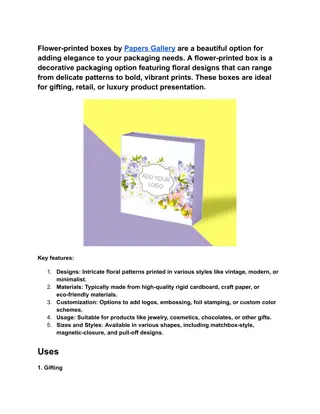
3D Printed Heads Enhancing ROSA Surgical Workflow
"Exploring the use of 3D-printed heads in SEEG and DBS surgeries at Beaumont Hospital through phantom design to improve surgical training, workflow refinement, and patient safety initiatives. The methodology involved creating head models from patients' pre-operative scans and utilizing them with ROSA for registration trials. Results show the potential benefits of using 3D-printed models in clinical teaching and training, leading to the establishment of curated workflows and skill development in robotic-assisted neurosurgery."

Download Presentation

Please find below an Image/Link to download the presentation.
The content on the website is provided AS IS for your information and personal use only. It may not be sold, licensed, or shared on other websites without obtaining consent from the author.If you encounter any issues during the download, it is possible that the publisher has removed the file from their server.
You are allowed to download the files provided on this website for personal or commercial use, subject to the condition that they are used lawfully. All files are the property of their respective owners.
The content on the website is provided AS IS for your information and personal use only. It may not be sold, licensed, or shared on other websites without obtaining consent from the author.
E N D
Presentation Transcript
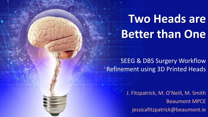
Two Heads are Better than One SEEG & DBS Surgery Workflow Refinement using 3D Printed Heads J. Fitzpatrick, M. O Neill, M. Smith Beaumont MPCE jessicafitzpatrick@beaumont.ie
Initiative & Aim In 2020, the ROSA One Brain was introduced into Beaumont Hospital for use in planning and performing Stereoelectroencephalography (SEEG), Deep Brain Stimulation (DBS), and biopsy procedures. The manufacturer is based in France; organising in-depth training was a challenge due to the Covid-19 pandemic with the intermittent closure of international borders. The neurophysics and neurosurgical team recognized a necessity to get familiar with ROSA workflows in a quick and safe manner but surgical opportunities and company support were limited. Figure 1: Zimmer Biomet ROSA Robot Aim: To explore the use of 3D printing as a means of developing imaging phantoms & replicate patient-specific features. Phantoms have the potential to test imaging modalities, processes and curate workflows in robotic-assisted neurosurgery and in this instance helped to facilitate the introduction of SEEG & DBS procedures to Beaumont Hospital
Methodology A head model was created from the first SEEG & DBS patients pre- operative scans and printed using a FDM printer. Both SEEG & DBS head models were hollow, with structures of interest included in the DBS print. The heads were used in conjunction with ROSA to trial the registration process using various registration techniques. Since each head model was based off a CT of that patient, a good correlation exists between the anatomy of the printed head and the imaging Figure 2: SEEG Head Model Figure 4: ROSA and the DBS Head During Pointer Registration Figure 3: Laser Alignment on Structures of Interest of DBS Head Model
Results & Conclusion This work is a foundation for further investigation of the potential for phantom design, materials and applications to support quality and patient safety initiatives. Using the head models allowed curated workflows for both SEEG and DBS to be established, which enabled the team to practice with ROSA as much as necessary before the actual surgery. Local expertise and skill was developed. It also demonstrates the potential for using 3D-printed models in clinical teaching and training. This avoided delays in the commencement of robotic SEEG and DBS surgery programmes which could have been affected by pandemic-related travel restrictions resulting in reduced availability of international on-site applications support. Acknowledgements Mr. Kieron Sweeney, Consultant Neurosurgeon Ms. Catherine Moran, Consultant Neurosurgeon Department of Materials and Manufacturing Engineering, DCU Representatives from ROSA & Brainlab Figure 5: DBS procedure underway using ROSA. (Credit: Laurent Vergeade, ROSA)