4.Bronchiectasis
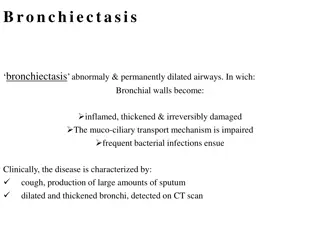
Bronchiectasis is a condition characterized by permanent dilation of bronchi and bronchioles due to chronic infections or obstructions. It presents with cough, expectoration of sputum, and may result from conditions like bronchial obstruction, necrotizing pneumonia, or chronic infections. The pathogenesis involves obstruction and chronic infection leading to irreversible airway dilation. Early diagnosis is crucial for appropriate management. Learn more about bronchiectasis in this informative content.

Download Presentation

Please find below an Image/Link to download the presentation.
The content on the website is provided AS IS for your information and personal use only. It may not be sold, licensed, or shared on other websites without obtaining consent from the author.If you encounter any issues during the download, it is possible that the publisher has removed the file from their server.
You are allowed to download the files provided on this website for personal or commercial use, subject to the condition that they are used lawfully. All files are the property of their respective owners.
The content on the website is provided AS IS for your information and personal use only. It may not be sold, licensed, or shared on other websites without obtaining consent from the author.
E N D
Presentation Transcript
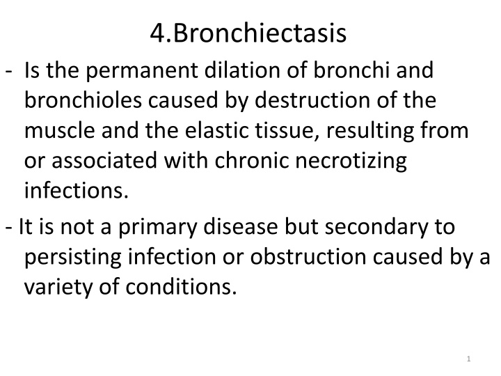
4.Bronchiectasis - Is the permanent dilation of bronchi and bronchioles caused by destruction of the muscle and the elastic tissue, resulting from or associated with chronic necrotizing infections. - It is not a primary disease but secondary to persisting infection or obstruction caused by a variety of conditions. 1
- Once developed, it gives rise to a characteristic symptom complex dominated by cough and expectoration of copious amounts of purulent sputum. - Diagnosis depends on an appropriate history along with radiographic demonstration of bronchial dilation. 2
The Predisposing conditions include: 1. Bronchial obstruction and common causes are : a- Tumors, foreign bodies, and impaction of mucus. - With these conditions, the bronchiectasis is localized to the obstructed lung segment. b- Bronchiectasis can also complicate atopic asthma and chronic bronchitis. 3
3.Necrotizing, or suppurative, pneumonia, particularly with Staphylococcus aureus or Klebsiella spp., may predispose affected patients to development of bronchiectasis. Note: Posttuberculosis bronchiectasis continues to be a significant cause of morbidity in endemic areas. 4
PATHOGENESIS of bronchiectasis: - Two processes are crucial in pathogenesis : obstruction and chronic infection and either of these may come first. - Normal clearance mechanisms are hampered by obstruction, so secondary infection soon follows conversely, chronic infection over time causes damage to ; bronchial walls, leading to weakening and dilation. 5
1- For example, obstruction caused by a primary lung cancer or a foreign body impairs clearance of secretions, providing substrate for superimposed infection and the resultant inflammatory damage to the bronchial wall and the accumulating exudate further distend the airways, leading to irreversible dilation. 6
2- Conversely, a persistent necrotizing inflammation in the bronchi or bronchioles may cause obstructive secretions, inflammation in the wall (with peribronchial fibrosis and traction on the walls), and eventually the train of events . 7
MORPHOLOGY - Bronchiectasis usually affects the lower lobes bilaterally, - When caused by tumors or foreign bodies the involvement may be localized to a single segment and the most severe involvement is in the more distal bronchi and bronchioles. 9
Gross:The airways may be dilated to as much as four times their usual diameter and on gross examination of the lung can be followed almost to the pleural surfaces - By contrast, in normal lungs, the bronchioles cannot be followed by ordinary gross examination beyond a point 2 to 3 cm from the pleural surfaces 10
Histologic findings 1. In the full-blown active case a. An intense acute and chronic inflammatory exudate within the walls of the bronchi and bronchioles b. Desquamation and ulceration of lining epithelium. c. a mixed flora can be cultured from the involved bronchi, 11
- When healing occurs, a. The lining epithelium may regenerate completely; however,usually so much injury has occurred that abnormal dilation and scarring persist. b- Fibrosis of the bronchial and bronchiolar walls and peribronchiolar fibrosis develop in more chronic cases.
c. In some instances, the necrosis destroys the bronchial or bronchiolar walls resulting in the formation of an abscess c avity within which a fungus ball may develop. 13
Clinical Features - Consist of severe, persistent cough with expectoration of mucopurulent, sometimes fetid, sputum. - The sputum may contain flecks of blood; frank hemoptysis can occur. . 14
- Symptoms often are episodic and are precipitated by upper respiratory tract infections or the introduction of new pathogenic agents. - Clubbing of the fingers may develop 15
Complications of bronchiectasis 1- In cases of severe, widespread bronchiectasis, significant obstructive ventilatory defects are usual, with hypoxemia, hypercapnia, pulmonary hypertension, and (rarely) cor pulmonale. 2- Metastatic brain abscesses 3- Reactive amyloidosis 16
Respiratory : restrictive lung diseases 17
1. Idiopathic pulmonary fibrosis (IPF), 18
- Also known as cryptogenic fibrosing alveolitis, refers to a pulmonary disorder of unknown etiology. - It is characterized by patchy but progressive bilateral interstitial fibrosis, which in advanced cases results in severe hypoxemia and cyanosis. 20
- Males are affected more often than females, - Two thirds of patients are older than 60 years of age at presentation. - The radiologic and histologic pattern of fibrosis is referred to as usual interstitial pneumonia (UIP), which is required for the diagnosis of IPF. 21
- However, similar pathologic changes in the lung may be present in well- defined entities such as asbestosis and the collagen vascular diseases. - Therefore, known causes must be ruled out before the term of idiopathic is used 22
PATHOGENESIS - IPF is caused by "repeated cycles" of epithelial activation/injury by unidentified agent - Histopathologic features include inflammation and induction of TH2 type T cell response with eosinophils, mast cells, IL-4, and IL-13 in the lesions. 23
- Alternatively activated macrophages are important in its pathogenesis - Abnormal epithelial repair at the sites of damage and inflammation gives rise to exuberant fibroblastic or myofibroblastic proliferation leading to the characteristic fibroblastic foci. 24
a.- TGF-1, which is released from injured type I pneumocytes induces transformation of fibroblasts into myofibroblasts leading to excessive and continuing deposition of collagen and ECM . b.- TGF- 1 also downregulates fibroblast caveolin-1, which acts as an endogenous inhibitor of pulmonary fibrosis 25
MORPHOLOGY - The pattern of fibrosis in IPF is referred to as usual interstitial pneumonia (UIP) -The histologic hallmark of UIP is patchy interstitial fibrosis, which varies in intensity and worsens with time. - The earliest lesions demonstrate exuberant fibroblastic proliferation and appear as fibroblastic Foci 26
Over time these areas become more collagenous and less cellular. - Quite typical is the existence of both early and late lesions (temporal heterogeneity) 27
- The dense fibrosis causes collapse of alveolar walls and formation of cystic spaces lined by hyperplastic type II pneumocytes or bronchiolar epithelium (honeycomb fibrosis). 30
- The interstitial inflammation usually is patchy and consists of an alveolar septal infiltrate of mostly lymphocytes and occasional plasma cells, mast cells, and eosinophils. 31
- Secondary pulmonary hypertensive changes (intimal fibrosis and medial thickening of pulmonary arteries) are often present. 32
2.Pneumoconioses - Is a term originally coined to describe the non-neoplastic lung reaction to inhalation of mineral dusts. - The term has been broadened to include diseases induced by organic as well as inorganic particulates . 33
- The mineral dust pneumoconioses- the three most common of which result from exposure to : a. Coal dust b. Silica, c. Asbestos 34
PATHOGENESIS : - The reaction of the lung to mineral dusts depends on their size shape, solubility, and reactivity -Effect of size a. 5 to 10 m Particles are unlikely to reach distal airways, b. Particles smaller than 0.5 m move into and out of alveoli, often without substantial deposition and injury. 35
c. 1 to 5 m particles are the most dangerous, because they get lodged at bifurcation of the distal airways. Reactivity 1- Coal dust is relatively inert, and large amounts must be deposited before lung disease is clinically detectable. 36
2. Silica, asbestos, and beryllium are more reactive than coal dust, resulting in fibrotic reactions at lower concentrations. Note: - Most inhaled dust is entrapped in the mucus blanket and rapidly removed from the lung by ciliary movement,- However, some of the particles become impacted at alveolar duct bifurcations, where macrophages accumulate and engulf the trapped particulates 37
- The alveolar macrophage is a key cellular element in the initiation and perpetuation of lung injury and fibrosis. a. Many particles activate the inflammasome and induce IL-1 b. The more reactive particles trigger the macrophages to 38
release a number of products that mediate inflammation and initiate fibroblast proliferation and collagen deposition. 39