Acute and Chronic Cholecystitis
Acute and chronic cholecystitis with cholelithiasis are comprehensively discussed by Prof. (Dr.) O.P. Gupta, highlighting causes, classification, mode of infection, pathogenesis, impacted stones, clinical presentation, and signs.

Download Presentation

Please find below an Image/Link to download the presentation.
The content on the website is provided AS IS for your information and personal use only. It may not be sold, licensed, or shared on other websites without obtaining consent from the author.If you encounter any issues during the download, it is possible that the publisher has removed the file from their server.
You are allowed to download the files provided on this website for personal or commercial use, subject to the condition that they are used lawfully. All files are the property of their respective owners.
The content on the website is provided AS IS for your information and personal use only. It may not be sold, licensed, or shared on other websites without obtaining consent from the author.
E N D
Presentation Transcript
Acute and Chronic Cholecystitis with Cholelithiasis Prof. (Dr.) O.P Gupta Professor & HOD Department of Surgery CIMS&H, Lucknow
Acute Cholecystitis Occurs in 1.Patients with pre existing chronic cholecystitis 2.As first episode Most Common Cause Impacted Gallstone in Hartmann s Pouch
ACUTE CHOLECYSTITIS Temporary impaction Only Biliary Colic No INFLAMMATION Prolonged impaction INFLAMMATION ENSURES Edema of GB Subserosal Hemorrhage
Causitive Organisms: E Coli (most common) Klebsiella,Pseudomonas,Proteus Strep.Faecalis Salmonella Clostridium Welchii
Classification Acute Calculous Cholecystitis Acute Acalculous Cholecystitis
Mode of Infection A. Hematogenous Hepatic Artery Cystic Artery A. Portal Vein B. Through Bile
Pathogenesis Stone causes Obstruction at hartmann s pouch or in cystic duct Obstruction causes Stasis It leads to Edema of the wall Bacterial Infection occurs Leads to Acute Cholecystitis
Impacted Stone -- mucosalerosion Thereby Bile Salts will act on Submucosal tissue Bile is toxic to tissues Leads to Necrosis, Infection and Perforation
Presentation SYMPTOMS FEVER RIGHT UPPER QUAD PAIN NAUSEA
SIGNS RIGHT UPPER QUAD TENDERNESS GUARDING RIGIDITY MURPHY S SIGN (arrest of inspiration with gentle pressure under the R costal margin due to tenderness) BOAA s SIGN- Hyperasthesia at 9th to 11th rib posteriorly on R side PALPABLE TENDER GALLBLADDER TACHYCARDIA
Predisposingfactors 1. Obesity 2. Female sex hormones estrogen & OCPs 3. Increasingage 4. Pregnancy 5. Drugs-octreotide,clofibrate 6. High fat diet 7. Diabetesmellitus
LITHOGENIC BILE STASIS OR HYPOMOTILITY OF GALL BLADDER NUCLEATION Increasecholesterol- obesity,diet Decrease bile acids- OCPs,genetic factors,PBC,ileal disease,ilealresection Excess pronucleating factors-e.g.mucin OCPs Vagotomy Fasting Pregnancy Prolonged parenteral nutrition Decreased anti- nucleating factors-e.g. Apolipoproteins Increase bilirubin- HemolyticAnemia
Types of Gallstones 1. Cholesterol stones radiating crystal like appearance 2. Mixed stones-Most common type ofstones; contains cholesterol, calcium salts of phosphatesand carbonates, palmitate ,proteins and are multiple faceted. 3. Pigment stones-small, black or greenish black, multiple and often sludgelike
Pigmentstones Black pigmentstones Mostcommon Formed in gallbladder Made of Calcium bilirubinate,phosphate, bicarbonate Common in hemolytic disorders, cirrhsis Multiple , small & hard in consistency Brown pigmentstones Rarely form in gall bladder Formed in bileduct Related to bile stasis& infected bile E.coli,Bacteroides
Clinicalfeatures More common infemales Fat,fertile,forty,flatulent 10% Gallstones are RADIO- OP AQUE Asymptomatic in 10 to 20%cases Symptoms- Biliary colic-Right hypochondrium & epigastrium, radiating to chest,back & shoulder, severe , on & off, spasmodic, occurs within hours after meal,usually self limiting and recurring,precipitatedby fattymeal. vomiting Fever IncreasedWBCs
INVESTIGATIONS LAB STUDIES LEUKOCYTOSIS Mild elevation of BILIRUBIN , ALP, SGOT/PT If Profound Jaundice + Picture of Acute Cholecystitis Suspect, CHOLANGITIS with obstruction of CBD MIRIZZI syndrome
USG Sensitive Inexpensive Reliable Sensitivity 85% and Specificity 95% What will you look in USG? 1.GallStone 2.Pericholecystic fluid 3.GB wall thickening 4.Sonographic murphy s sign
Accurate History Physical Examination Supportive Lab Studies And an Ultrasound Needed in most of the cases for Diagnosis Extras HIDAScan CT Scan
Remember CT is less sensitive than USG for the diagnosis of Acute Cholecystitis
Management of Acute Cholecystitis 1. NPO 2. RYLES TUBE ASPIRATE 3. IV Fluids 4.BROAD SPECTRUM ANTIBIOTICS 5.IV ANALGESICS 6. OBSERVATION
Surgery in a/c Cholecystitis When presents within 2 to 3 days LAP CHOLECYSTECTOMY When presents more than 3 days INTERVAL CHOLECYSTECTOMY after 6 weeks In Pregnancy CHOLECYSTECTOMY @ T2 Empyema, Persisting and Progressing Symptoms CHOLECYSTECTOMY EMERGENCY
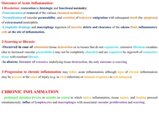
Complications PERFORATION PERITONITIS PERICHOLECYSTIC ABSCESS CHOLANGITIS and SEPTICEMIA PANCREATITIS EMPYEMA GB GANGRENOUS GB
Acute Acalculous Cholecystitis 5% ICU patients, Post OP patients, Burns, Cholecystoses Gallbladder distension, release of Factor VII Acute Presentation Rx Cholecystectomy
Chronic Cholecystitis Chronically Inflammed Thickened Gallbladder which is NONFunctioning NONdistending
Causes GALL STONES CHOLECYSTOSIS CHRONIC ACALCULOUS CHOLECYSTITIS Organisms: Klebsiella Steptococci Salmonella
Pathology GB is shrunken, contracted,small, nonfunctioning, fibrotic with thickened GB wall Mucosa proliferates into Lumen ROKITANSKY ASHCHOFF SINUSES Muscular wall replaced by Fibrotic tissue
Clinical Featues Colicky Pain Murphy s Sign Dyspeptic Symptoms Intolerance to fatty meal
Complications of Gallstones In Bileduct- InIntestine- In GallBladder- Acutecholecystitis Chroniccholecystitis Empyema of gallbladder Mucocele gallbladder Perforation leading to biliary peritonitis Gangrene of gallbladder Carcinoma Obstructive jaundice Acuteintestinal obstruction Cholangitis Acute pancreatitis
Treatment of Chronic Cholecystitis is CHOLECYSTECTOMY
Surgicaltherapy Laparoscopic cholecystectomy is ideal. Open cholecystectomy is done if patient unfit for laparoscopy through Right Sub-costal (KOCHERS s) incision.