Acute Bacterial Meningitis

Bacterial meningitis is an acute purulent infection within the subarachnoid space, involving the meninges, subarachnoid space, and brain parenchyma. Learn about predisposing factors, etiology, pathogenesis, clinical presentation, and signs of meningeal irritation associated with this condition.

Download Presentation

Please find below an Image/Link to download the presentation.
The content on the website is provided AS IS for your information and personal use only. It may not be sold, licensed, or shared on other websites without obtaining consent from the author.If you encounter any issues during the download, it is possible that the publisher has removed the file from their server.
You are allowed to download the files provided on this website for personal or commercial use, subject to the condition that they are used lawfully. All files are the property of their respective owners.
The content on the website is provided AS IS for your information and personal use only. It may not be sold, licensed, or shared on other websites without obtaining consent from the author.
E N D
Presentation Transcript
Bacterial meningitis is an acute purulent infection within the subarachnoid space. The meninges, the subarachnoid space, and the brain parenchyma are all frequently involved in the inflammatory reaction (meningoencephalitis). PREDISPOSING FACTORS: Systemic(especially respiratory ) or parameningeal infections DM, alcoholism, cancer, or other immunodeficiency states. Anatomical meningeal defect, prior neurosurgery, or head trauma.
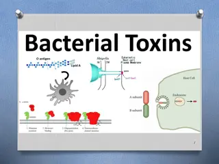
ETIOLOGY S. pneumoniae (pneumococcal meningitis) is the most common cause account for 50% of all cases of bacterial meningitis mainly in adults >20 years of age. N. meningitidis (meningococcal meningitis) accounts for 25% of all cases of meningitis mostly in children and young adults between the ages of 2 and 20. Group B streptococcus, or S. agalactiae: accounts for 15% of meningitis predominantly in neonates. L. monocytogenes accounts for 10% predominantly in neonates (<1 month of age), pregnant women, individuals >60 years, and immunocompromised individuals of all ages. H. influenzae type b accounts for 10% of bacterial meningitis mainly in children. Staphylococcus aureus are important causes of meningitis that occurs following invasive neurosurgical procedures, particularly shunting procedures, or as a complication of of intrathecal chemotherapy.
PATHOGENESIS Bacteria nasopharynx,leading to tissue invasion and hematogenous spread to the SAS. typically gain access to the CNS by colonizing the Bacteria can also spread to the meninges directly, through anatomic defect in the skull or from parameningeal sites such as paranasal sinuses or middle ear. The low level of antibodies and complement and the few WBCs in the CSF are inadequate to contain the infection. The resulting inflammatory response resulting in release of cytokines that promote BBB permeability ,vasogenic edema, changes in cerebral blood flow and perhaps direct neuronal toxicity
CLINICALPRESENTATION Presentation is usually acute and patient may have meningitis for 1-7 days . symptoms of The classic clinical triad of meningitis is fever, headache, and nuchal rigidity. A decreased level of consciousness occurs in >75% of patients and can vary from lethargy to coma. Nausea, vomiting, and photophobia are also common complaints. Seizures occur as part of the initial presentation of bacterial meningitis or during the course of the illness in 20 40% of patients.
Signs of meningeal irritation are seen in approximately 80% of cases, (often absent in the very young and very old, or with profound impairment of consciousness). These include: neck stiffness on passive flexion, thigh flexion upon flexion of the neck brudzinski sign , resistance to passive extension of the knee with the hip flexed kernig sign. A petechial rash is seen in 50-60% of patients with N.meningitidis meningitis
Blood Studies complete blood count (CBC) with differential will demonstrate polymorphonuclear leukocytosis. Serum electrolytes, to determine dehydration or syndrome of inappropriate secretion of antidiuretic hormone (SIADH) Serum glucose (which is compared with the CSF glucose) Blood urea nitrogen (BUN) or creatinine and liver profile, to assess organ function and adjust antibiotic dosing
Cultures and Bacterial Antigen Testing The utility of cultures is most evident when LP is delayed until head imaging can rule out the risk of brain herniation, in which cases antimicrobial therapy is rightfully initiated before CSF samples can be obtained. These cultures include the following: Blood - 50% positive in meningitis caused by H influenzae, S pneumoniae, or N meningitidis Nasopharynx Respiratory secretions Urine Skin lesions
DIAGNOSIS The diagnosis of bacterial meningitis is made by examination of the CSF . The need to obtain neuroimaging studies (CT or MRI) prior to LP requires clinical judgment. In an immunocompetent patient with no history of head trauma, a normal level of consciousness, and no papilledema or focal neurologic deficits, it is safe to perform LP without prior neuroimaging studies.
The classic CSF abnormalities in bacterial meningitis are: (1) polymorphonuclear (PMN) leukocytosis (>100 cells/L ). (2) decreased glucose concentration (<40 mg/dL) and/or CSF/serum glucose ratio of <0.4 (3) increased protein concentration (>45 mg/dL) . (4) increased opening pressure (>180 mmH2O ). CSF bacterial cultures are positive in >80% of patients, and CSF Gram's stain demonstrates organisms in >60%.
broad-based bacterial PCR can detect small numbers of viable and nonviable organisms in CSF and is expected to be useful for making a diagnosis of bacterial meningitis in: patients who have been pretreated with oral or parenteral antibiotics in whom Gram's stain and CSF culture are negative. When the broad-range PCR is positive, a PCR that uses specific bacterial primers to detect the nucleic acid of S. pneumoniae, N. meningitidis, Escherichia coli, L. monocytogenes, H. influenzae, and S. agalactiae can be obtained based on the clinical suspicion of the meningeal pathogen.
The latex agglutination (LA) test for the detection of bacterial antigens of S. pneumoniae, N. meningitidis, H. influenzae type b, group B streptococcus, and E. coli strains in the CSF has been useful for making a diagnosis of bacterial meningitis but is being replaced by the CSF bacterial PCR assay. The CSF LA test has a specificity of 95 100% for S. pneumoniae and N. meningitidis, so a positive test is virtually diagnostic of bacterial meningitis caused by these organisms. However, the sensitivity of the CSF LA test is only 70 100% for detection of S. pneumoniae and 33 70% for detection of N. meningitidis antigens, so a negative test does not exclude infection by these organisms.
DIFFERNTIAL DIAGNOSIS Viral encephalitis, can mimic the clinical presentation of bacterial meningitis. The findings on CSF studies, neuroimaging, and electroencephalogram (EEG) distinguish HSV encephalitis from bacterial meningitis. meningoencephalitis, and particularly herpes simplex virus (HSV) Focal CNS infections , including subdural and epidural empyema and brain abscess, should also be considered, especially when focal neurologic findings are present. MRI should be performed in all patients with suspected meningitis who have focal features. subacutely evolving meningitis may be considered in the differential diagnosis of acute meningitis. The principal causes include Mycobacterium tuberculosis ,and Brucelosis. A number of noninfectious CNS disorders can mimic bacterial meningitis. Subarachnoid hemorrhage SAH is generally the major consideration. Other possibilities include chemical meningitis due to rupture of tumor contents into the CSF (e.g., from a cystic glioma or craniopharyngioma epidermoid or dermoid cyst);
TREATMENT Bacterial meningitis is a medical emergency. The goal is to begin antibiotic therapy within 60 min of a patient's arrival in the emergency room. Antimicrobial therapy is initiated in patients with suspected bacterial meningitis before the results of CSF Gram's stain and culture and after obtaining blood sample for culture. The initial choice of antibiotics is empirical , based upon the age of the patient and the predisposing factors .therapy is adjusted as indicated when the gram stain or culture and sensitivity result become available Lumber puncture can be repeated to assess the response to therapy.CSF should be sterile after 24 hours, and decrease in pleocytosis should occur within 3 days ,while sugar may remain low for many days . Dexamethasone ,given immediately before the onset of antibiotics treatment and continued for 4 days, can reduce mortality in patient with meningitis
Antibiotics Used in Empirical Therapy of Bacterial Meningitis Indication Less than 3 months Ampicillin + cefotaxime or ceftriaxone Indication Antibiotic Antibiotic alternative agent chloramphenicol + gentamicin 3months -50 years Cefotaxime , ceftriaxone or cefepime + vancomycin chloramphenicol + vancomycin Or clindamycin or meropenem Adults >55 and adults of any age with alcoholism or other debilitating illnesses Ampicillin + cefotaxime, ceftriaxone or cefepime + vancomycin meropenem, TMP-SMX, or doxycycline. Hospital-acquired meningitis, posttraumatic or postneurosurgery meningitis, neutropenic patients, or patients with impaired cell-mediated immunity Ampicillin + ceftazidime + vancomycin meropenem + vancomycin
Child (>1 month) Adult Ampicillin 200 (mg/kg)/d, q4h 12 g/d, q4h Cefepime 150 (mg/kg)/d, q8h 6 g/d, q8h Cefotaxime 200 (mg/kg)/d, q6h 12 g/d, q4h Ceftriaxone 100 (mg/kg)/d, q12h 4 g/d, q12h Ceftazidime 150 (mg/kg)/d, q8h 6 g/d, q8h Gentamicin 7.5 (mg/kg)/d, q8hb 7.5 (mg/kg)/d, q8h Meropenem 120 (mg/kg)/d, q8h 3 g/d, q8h Metronidazole 30 (mg/kg)/d, q6h 1500 2000 mg/d, q6h Nafcillin 100 200 (mg/kg)/d, q6h 9 12 g/d, q4h Penicillin G 400,000 (U/kg)/d, q4h 20 24 million U/d, q4h Vancomycin 60 (mg/kg)/d, q6h 2 g/d, q12hb
PROGNOSIS Mortality is 20% and the risk of death from bacterial meningitis increases with : (1) Decreased level of consciousness on admission. (2) Onset of seizures within 24 h of admission. (3) Signs of increased ICP (4) Young age (infancy) and age >50 (5) The presence of comorbid conditions including shock and/or the need for mechanical ventilation. (6) Delay in the initiation of treatment. Common sequelae include: decreased intellectual function, memory impairment, headache, hydrocephalus, SIADH, seizures, hearing loss and dizziness, and gait disturbances.
Prevention Vaccinations are available against pneumococci, meningococci, and hemophilus influenzae type b (Hib) and are recommended in high risk children. All contacts with patients (individuals who have had contact with oropharyngeal secretions, either through kissing or by sharing toys, beverages, or cigarettes). meningococcal meningitis should be given prophylactic rifampicin at a dosage of 10 mg/kg in children and 600 mg/day in adults for 2 days. In case of any contraindication for the use of rifampicin, asingle intramuscular injection of ceftriaxone may be given.
ETIOLOGY COMMON COMMON LESS COMMON LESS COMMON Enteroviruses echoviruses,coxsackiviruses, and human enteroviruses VZV HSV-2 Arboviruses EBV
CLINICAL MANIFESTATIONS Headache : usually frontal or retro- orbital and is often associated with photophobia and pain on moving the eye . Nuchal rigidity :present in most cases ,but may be mild and present only near the limit of neck anteflexion. Constitutional symptoms : fever, malaise, myalgia ,nausea and vomiting, abdominal pain and/or diarrhea. Patient may have mild lethargy or drowsiness. Sings of meningeal irritations: neck stiffness, Kernigs sign and Brubzinskis sign
LABORATORY DIAGNOSIS CSF Examination is the most important laboratory test in the diagnosis of viral meningitis . the typical profile is; WBC: (25 500 cells/mL) predominantly lymphocytes. Protein: normal or slightly elevated (20 80 mg/dl). Glucose : normal. Opening pressure : normal or slightly elevated (100 350 mm H2O). Polymerase Chain Reaction Amplification of viral specific DNA or RNA from CSF using PCR amplification has become the single most important method for diagnosing CNS viral infections . Viral Culture The sensitivity of CSF cultures for the diagnosis of viral meningitis and encephalitis, in contrast to it utility in bacterial infection ,is generally poor.
Serological Studies Serum antibodies determination remain a crucial diagnostic test for viruses with low seroprevalence rates in the general population. Neuroimaging Study MRI and CT scan are not necessary for patients with uncomplicated viral meningitis ,but should be performed in patients with altered consciousness, seizures, focal neurological sings or symptoms ,or atypical CSF profiles.
TREATMENT Treatment of all patients is primarily symptomatic and includes use of analgesics, antipyretics, antiemetics with fluid and electrolytes monitoring . Hospitalization is required for those patients who are immunocompromised; have significant alteration of consciousness, seizures, or the presence of focal signs and symptoms suggesting parenchymal brain involvement ; and those who have atypical CSF profile Oral or intravenous acyclovir may be of benefit in patients with meningitis caused by HSV, severe EBV or VZV infection. Seriously ill patients should receive intravenous acyclovir (15 30 mg/kg per day in three divided doses) which can be followed by an oral drug such as acyclovir (800 mg five times daily),famciclovir (500 mg tid), or valacyclovir (1000 mg tid) for a total course of 7 14 days. Patients who are less ill can be treated with oral drug alone .
PREVENTION Vaccination is an effective method of preventing the development of meningitis and other neurological complications associated with poliovirus, mumps, and measles . PROGNOSIS In adult the prognosis for full recovery from viral meningitis is excellent. Rare patients complain of persistent headache, incoordination, or generalized asthenia for weeks to months . mild mental impairment,
In contrast to viral meningitis, where the infectious process and associated inflammatory response are limited largely to the meninges, in encephalitis the brain parenchyma is also involved. Many patients with encephalitis also have evidence of associated meningitis (meningoencephalitis).
ETIOLOGY COMMON LESS COMMON Herpesviruses HSV-1 VZV EBV Rabies Estern equine virus Western equine virus CMV Enteroviruses Mumps Arthropod-born viruses
CLINICALMANIFESTATIONS In addition to the acute febrile illness with evidence of meningeal involvement characteristic of meningitis, the patient with encephalitis commonly has : depressed level of consciousness, ranging from mild lethargy to coma. evidence of focal weakness, involuntary movements, and cranial nerves deficits). neurologic signs and symptoms (aphasia, ataxia, Patients may also have confusion, hallucinations, agitation, behavioral disorders, and, at times, a frank psychosis. Focal or generalized seizures may occur.
Laboratory Diagnosis CSF Examination CSF examination should be performed in all patients with suspected viral encephalitis unless contraindicated by the presence of severely increased ICP. The characteristic CSF profile is indistinguishable from that of viral meningitis. CSF PCR CSF Culture Serologic Studies and Antigen Detection MRI, CT, EEG Patients with suspected encephalitis almost invariably undergo neuroimaging studies and often EEG. These tests help identify or exclude alternative diagnoses and assist in the differentiation between a focal, as opposed to a diffuse, encephalitic process. Focal findings in a patient with encephalitis should always raise the possibility of HSV encephalitis which usually affect the frontotemporal surface of the brain. Brain Biopsy
Treatment many patients will require care in an intensive care unit with monitoring of the vital functions, careful monitoring of ICP, fluid restriction, and suppression of fever. Seizures should be treated with standard anticonvulsant regimens, and prophylactic therapy should be considered. As with all seriously ill, immobilized patients, encephalitis patients are at risk for aspiration pneumonia, bed sores, DVT, and infections of indwelling lines and catheters. Acyclovir is of benefit in the treatment of HSV and should be started empirically in patients with suspected viral encephalitis, especially if focal features are present . Adults should receive a dose of 10 mg/kg of acyclovir intravenously every 8 h for a minimum of 14 days.
ACYCLOVIR Prior to intravenous administration, acyclovir should be diluted to a concentration 7 mg/mL. (A 70-kg person would receive a dose of 700 mg, which would be diluted in a volume of 100 mL.) Each dose should be infused slowly over 1 h rather than by rapid or bolus infusion, to minimize the risk of renal dysfunction. Care should be taken to avoid extravasation or intramuscular or subcutaneous administration. The alkaline pH of acyclovir can cause local inflammation and phlebitis Complications of therapy include elevations in blood urea nitrogen and creatinine levels , thrombocytopenia , gastrointestinal toxicity (nausea, vomiting, diarrhea) , and neurotoxicity (lethargy , disorientation, confusion, agitation, hallucinations, tremors, seizures) .
PROGNOSIS The incidence and severity of sequelae were directly related to the age of the patient and the level of consciousness at the time of initiation of therapy. Patients with severe neurologic impairment (Glasgow coma score 6) at initiation of therapy either died or survived with severe sequelae. Young patients (<30 years) with good neurologic function at initiation of therapy did substantially better (100% survival, 62% with no or mild sequelae) compared with their older counterparts (>30 years; 64% survival, 57% no or mild sequelae).
pathopysiology Tuberculous meningitis in adults does not develop acutely from hematogenous spread of tubercle bacilli to the meninges. Rather,( miliary) tubercles form in the parenchyma of the brain during hematogenous dissemination of tubercle bacilli in the course of primary infection. These tubercles enlarge and are usually caseating. Subependymal caseous foci cause meningitis via discharge of bacilli and tuberculous antigens into the SAS, producing an intense inflammatory reaction that leads to the production of a thick exudate that fills the basilar cisterns and surrounds the cranial nerves and major blood vessels at the base of the brain.
Clinical Manifestations Patients with subacute meningitis typically have headache, stiff neck, low- grade fever, night sweats and lethargy for days to several weeks before presentation. The meningitis is often concentrated around the base of the brain, causing cranial nerve palsies. Elevated intracranial pressure and hydrocephalus are common. It can also cause inflammation of the blood vessels as they leave the meninges to enter the brain, causing strokes.
Laboratory Diagnosis The classic CSF abnormalities in tuberculous meningitis are as follows: (1) elevated opening pressure. (2) lymphocytic pleocytosis (10 500 cells/L). (3) elevated protein concentration in the range of 1 5 g/L (10 500 mg/dL). (4) decreased glucose concentration in the range of 1.1 2.2 mmol/L (20 40 mg/dL). The combination of unrelenting headache, stiff neck, fatigue, night sweats, and fever with a CSF lymphocytic pleocytosis and a mildly decreased glucose concentration is highly suspicious for tuberculous meningitis.
Positive smears are typically reported in only 1040% of cases of tuberculous meningitis in adults. Cultures of CSF take 4 8 weeks to identify the organism and are positive in ~50% of adults. Culture remains the "gold standard" to make the diagnosis of tuberculous meningitis. PCR for the detection of M. tuberculosis DNA has a sensitivity of 70 80% but is limited at the present time by a high rate of false-positive results.
Treatment Empirical therapy of tuberculous meningitis is often initiated on the basis of a high index of suspicion without adequate laboratory support. first 2 months of intensive treatment require quadruple therapy with the combination of : isoniazid (300 mg/d), rifampin (10 mg/kg per day), pyrazinamide (30 mg/kg per day in divided doses), with the addition of one of the following: ethambutol (15 20mg/kg/day), streptomycin (15 mg/kg/day; maximum 1 g), ethionamide (15 20 mg/kg/day). Isoniazid which has excellent CSF penetration should be given together with rifampicin for the continuation phase of treatment from months 3 to 9. Pyridoxine therapy (100 mg/day) should be given with isoniazid therapy to prevent the development of peripheral neuropathy. Adjunctive corticosteroids, significantly reduce mortality across all severity grades. For this reason, steroids should be administered in all cases from day 1 of treatment, in a tapering dose for the first 1 2 months.
Definition A brain abscess is a focal, suppurative infection within the brain parenchyma, typically surrounded by a vascularized capsule. The term cerebritis is often employed to describe a nonencapsulated brain abscess. Etiology Abrain abscess may develop (1) by direct spread from a contiguous cranial site of infection, such as paranasal sinusitis, otitis media, mastoiditis, or dental infection. (2) following head trauma or a neurosurgical procedure. (3) as a result of hematogenous spread from a remote site of infection. The most important pathogens are: Streptococcus spp. (40%), staphylococci (10%). Enterobacteriaceae. (25%), anaerobes (30%), and
Clinical Presentation The most common symptoms in patients with a brain abscess are : headache, often characterized as a constant, dull, aching sensation, either hemicranial or generalized, and it becomes progressively more severe and refractory to therapy. Fever is present in only 50% of patients at the time of diagnosis, and its absence should not exclude the diagnosis. focal or generalized seizures. Focal neurologic deficits including hemiparesis, aphasia, or visual field defects . Signs of raised ICP papilledema, nausea and vomiting. Meningismus is not present unless the abscess has ruptured into the ventricle or the infection has spread to the subarachnoid space.
Diagnosis Diagnosis is made by neuroimaging studies. MRI is better than CT for demonstrating abscesses in the early (cerebritis) stages and is superior to CT for identifying abscesses in the posterior fossa. Microbiologic diagnosis of the etiologic agent is most accurately determined by Gram's stain and culture of abscess material obtained by stereotactic needle aspiration. LP should not be performed in patients with known or suspected focal intracranial infections. Additional laboratory studies may provide clues to the diagnosis of brain abscess in patients with a CNS mass lesion. About 50% of patients have a peripheral leukocytosis, 60% an elevated ESR, and 80% an elevated C- reactive protein. Blood cultures are positive in ~10% of cases
Differential Diagnosis Conditions that can cause headache, seizure activity include: brain abscess, meningoencephalitis, disseminated encephalomyelitis. fever, focal neurologic signs, and subdural empyema, superior sagittal sinus thrombosis, bacterial meningitis, viral and acute When fever is absent: primary and metastatic brain tumors become the major differential diagnosis. Less commonly, cerebral infarction or hematoma can have an MRI or CT appearance resembling brain abscess.
Treatment Optimal therapy of brain abscesses involves a combination of high-dose parenteral antibiotics and neurosurgical drainage. Empirical therapy of brain abscess typically includes a third-generation cephalosporin (e.g., cefotaxime or ceftriaxone) and metronidazole. In patients with head trauma or neurosurgical procedures, treatment should include ceftazidime as the third-generation cephalosporin to enhance coverage of Pseudomonas spp. and vancomycin for coverage of staphylococci. Intravenous dexamethasone therapy (10 mg every 6 h) is usually reserved for patients with substantial periabscess edema and associated mass effect and increased ICP.
Aspiration and drainage of the abscess under stereotactic guidance are beneficial for both diagnosis and therapy, but complete excision of a bacterial abscess via craniotomy or craniectomy is generally reserved for multiloculated abscesses or those in which stereotactic aspiration is unsuccessful. Serial MRI or CT scans should be obtained on a monthly or twice- monthly basis to document resolution of the abscess


 test for the detection of")


")