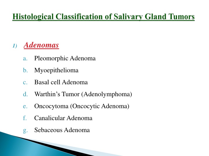
Adenomas
The diverse types of tumors affecting salivary glands, including adenomas, carcinomas, non-epithelial tumors, malignant lymphomas, secondary tumors, and unclassified tumors. Learn about specific subtypes like pleomorphic adenoma, myoepithelioma, acinic cell carcinoma, and more. Discover the clinical features, common sites, and cellular differentiation processes involved in these tumors.

Download Presentation

Please find below an Image/Link to download the presentation.
The content on the website is provided AS IS for your information and personal use only. It may not be sold, licensed, or shared on other websites without obtaining consent from the author.If you encounter any issues during the download, it is possible that the publisher has removed the file from their server.
You are allowed to download the files provided on this website for personal or commercial use, subject to the condition that they are used lawfully. All files are the property of their respective owners.
The content on the website is provided AS IS for your information and personal use only. It may not be sold, licensed, or shared on other websites without obtaining consent from the author.
E N D
Presentation Transcript
1) Adenomas a. PleomorphicAdenoma b. Myoepithelioma c. Basal cell Adenoma d. Warthin s Tumor (Adenolymphoma) e. Oncocytoma (OncocyticAdenoma) f. Canalicular Adenoma g. Sebaceous Adenoma
h.Ductal Papilloma : a. Inverted Ductal Papilloma b. Intraductal Papilloma c. Sialadenoma Papilleferum i. Cystadenoma a. Papillary cystadenoma b. Mucinous Cystadenoma
2) Carcinomas a. Acinic cell carcinoma b. Mucoepidermoid carcinoma c. Adenoid cystic carcinoma d. Polymorphous low grade adenocarcinoma (Terminal duct Adenocarcinoma) e. Epithelial myoepithelial carcinoma
f. Basal cell adenocarcinoma g. Sebaceous carcinoma h. Papillary Cystadenocarcinoma i. Mucinous Cystadenocarcinoma j. Oncocytic Carcinoma k. Salivary duct Carcinoma l. Adenocarcinoma
m. Malignant myoepithelioma (Myoepithelial carcinoma) n. Carcinoma in pleomorphic adenoma ( Malignant mixed tumor) o. Squamous cell carcinoma p. Small cell carcinoma q. Undifferentiated carcinoma r. Other carcinomas
3) Non Epithelial Tumors 4) Malignant Lymphomas 5) Secondary Tumors 6) Unclassified Tumors 7) Tumor Like Lesions: a. Sialadenosis b. Oncocytosis c. Necrotizing Sialometaplasia d. Benign Lymphoepithelial tumors e. Salivary Gland cyst f. Chronic sclerosing sialadenitis of submandibular gland (Kuttner tumor) g. Cystic Lymphoid hyperplasia in AIDS
Most common 53-77% - Parotid gland 44- 68% - Submandibular gland 38- 43% - Minor glands Derived from a mixture of ductal and myoepithelial elements
That is reserve cells in intercalated duct Intercalated duct reserve cell differentiate into ductal and myoepithelial cell Myoepithelial cell can undergo mesenchymal metaplasia and gives rise to morphologic diversity of this tumor Not truly a mixed neoplasm
Clinical Features Parotid gland is most common site 4thto 6thdecade ,female predilection i.e. 6:4 Also common in young adults, children. Small, painless quiescent nodule Slowly grow and increase in size Sometimes show intermittent growth
Do not show fixation to deeper tissue or the overlying skin It is an irregular nodular lesion that is firm in consistency. Areas of cystic degeneration may be palpated if superficial. Skin seldom ulcerates. Pain is not a common symptom. Local discomfort is frequently present
Most pleomorphic adenomas of parotid gland occur in the superficial lobe It presents as a swelling overlying the mandibular ramous in front of ear Facial palsy in rare Tumor initially is mobile, but becomes less mobile as it grows larger
Intraoral Tumors < 1- 2 cm in diameter Palate is the most common site for minor gland followed by upper lip and buccal mucosa Causes difficulty in mastication, talking and breathing Palatal Tumors on posterior lateral aspect Present as smooth surfaced, dome shaped mass If traumatized ulcerates Appear fixed to underlying bone but is noninvasive
Histopathology Well circumscribed encapsulated tumor Capsule may be incomplete/ show infiltration Commonly seen in minor gland tumors tumor is composed of mixture of glandular epithelium and myoepithelial cells in mesenchyme like background
The epithelium often forms ducts and cystic structures or may from islands or sheets of cell. Keratinizing sq. cells i.e. mucous producing cells can also be seen. Myoepithelial Cells Present in large number Have variable morphology angular/ Spindle
Some are rounded ; eccentric nucleus and hyalinized eosinophilic cytoplasm; resembling plasma cell. Characteristically; plasmacytoid appearance of myoepithelial cell is seen in minor gland tumors. Stromal change is produced by the myoepithelial cells. Collection of mucoid material in tumor cells result in myxomatous background
Vacuolar degeneration of cells produce chondroid appearance. At places eosinophilic hyalinized change At times, fibrous, fat or osteoid is seen Presence of entirely myoepithelial cell as myoepitheliomas
Benign Salivary Gland Tumors Benign Salivary Gland Tumors
Treatment Surgical Excision In Parotid The tumor and involved lobe of the gland is removed. Submax gland tumors are treated by removal of gland and tumor in continuity Intraoral by extracapsular excision Palatal tumor- excised overlying mucosa Lining mucosa enucleation/ extracapsular excision
 Adenomas")

 Carcinomas")


 Non Epithelial Tumors")