Advanced Obstetrics Ultrasound Scanning Images
Explore a series of detailed ultrasound images showcasing the development and measurements of a fetus during the first and second trimesters of pregnancy. Witness key stages such as fetal pole and embryo formation, fetal viability with heart rate detection, crown-rump length measurement, nuchal translucency assessment, and more.

Download Presentation

Please find below an Image/Link to download the presentation.
The content on the website is provided AS IS for your information and personal use only. It may not be sold, licensed, or shared on other websites without obtaining consent from the author. If you encounter any issues during the download, it is possible that the publisher has removed the file from their server.
You are allowed to download the files provided on this website for personal or commercial use, subject to the condition that they are used lawfully. All files are the property of their respective owners.
The content on the website is provided AS IS for your information and personal use only. It may not be sold, licensed, or shared on other websites without obtaining consent from the author.
E N D
Presentation Transcript
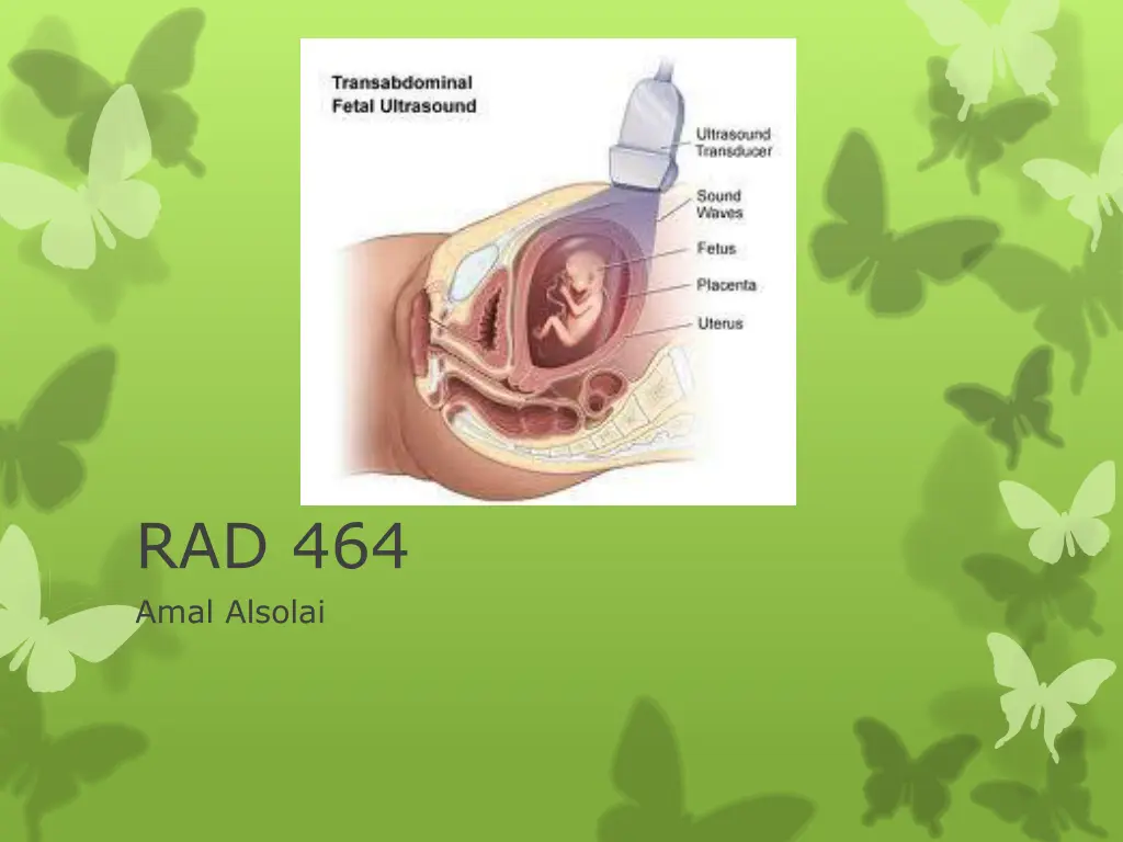
RAD 464 Amal Alsolai
Obstetrics U/S 1 /First Trimester Scanning 6 wks TO 12 wks Scans
Sac with Embryo>CRL, yolk sac ,aminion
NT Measurment< nuchal translucency 10 to 14 weeks
SECOND TRIMESTER SCANNING 18 TO 40 WEEKS
2/SECOND Trimester Scanning Fetal Measuments *Head circumferance