Alteration in Hematologic Function Lecture 7.1 Overview
Introduction to the physical characteristics, components, volume, functions, and percentages of blood, focusing on plasma and formed elements like red blood cells, white blood cells, and platelets. Details on blood plasma composition, erythrocytes, and hematopoiesis provided.

Download Presentation

Please find below an Image/Link to download the presentation.
The content on the website is provided AS IS for your information and personal use only. It may not be sold, licensed, or shared on other websites without obtaining consent from the author.If you encounter any issues during the download, it is possible that the publisher has removed the file from their server.
You are allowed to download the files provided on this website for personal or commercial use, subject to the condition that they are used lawfully. All files are the property of their respective owners.
The content on the website is provided AS IS for your information and personal use only. It may not be sold, licensed, or shared on other websites without obtaining consent from the author.
E N D
Presentation Transcript
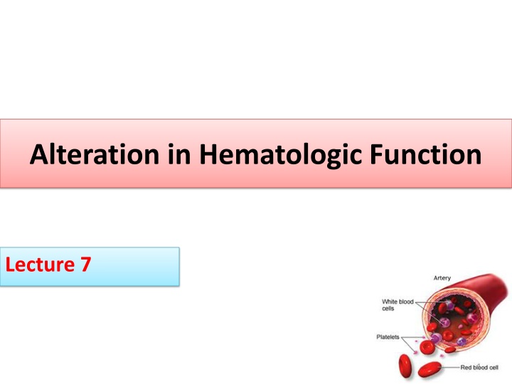
Alteration in Hematologic Function Lecture 7 1
Introduction Artery White blood cells Platelets Red blood cells 2
Physical Characteristics of Blood Heavier, thicker, and 3-4 more viscous than water 38o C (100.4oF) pH : 7.35 7.45 4-6 liters in an adult. Varies with electrolyte concentration and amount of adipose tissue 3
Components of the Blood Blood: Plasma water, albumin, electrolytes, clotting factors Cellular Components RBCs, WBCs, Platelets All formed in the red bone marrow (after birth) In utero- spleen, thymus, liver lymphatic system regulates maturation
Blood Volume Blood volume is about 8% of body weight. 1 kg of blood 1 L of blood 70 kg X 0.08 = 5.6 Kg = 5.6 L 45 % is formed elements 55% plasma 5
Blood Functions 1. Deliver O2 2. Remove metabolic wastes 3. Maintain temperature, pH, and fluid volume 4. Protection from blood loss- platelets 5. Prevent infection- antibodies and WBC 6. Transport hormones 6
Blood Percentages 55 % plasma Plasma is the straw-colored liquid in which the blood cells are suspended. 45 % formed elements Red blood cells (Erythrocytes) White blood cells (leukocytes) Platelets (thrombocytes) 7
Blood Plasma-55% Buffy coat-<1% Formed elements-45% 8
Plasma Plasma consists of: 90% water. 10 % solutes: albumin, electrolytes and proteins. Proteins consist of clotting factors, globulins, circulating antibodies and fibrinogen. 10
Erythrocytes RBCs carry hemoglobin which is attached to oxygen- provides O2 to the tissues life span 120 days manufacture regulated by erythropoetin Normal Hematocrit- 35-45% Normal Hemoglobin- 12-16 grams Hematopoiesis- production of RBC. Polycethemia: increase the number of RBC
RBCs The bones are continually producing new cells. 12
RBC breakdown Healthy RBC s live about 120 days; we break down about 174 million per minute RBC s are removed from circulation by the liver and spleen Broken down into heme and globin portions Globin is broken down into amino acids Iron is removed from heme and stored or recycled Heme is broken down into biliverdin and then into bilirubin 13
RBC breakdown Usually eliminated in bile. To produce more RBC s, the body needs sufficient iron and amino acids as well as the vitamins folate (folic acid) and vitamin B12 Erythropoietin is a hormone produced by the kidneys in response to low blood oxygen levels; signals bone marrow to increase RBC production. 14
Leukocytes: White Blood Cells The battling blood cells. The white blood cells are continually on the look out for signs of disease. They are five types Leukopenia: decrease NO of WBC When a germ appears the WBC will: Produce protective antibodies. Surround it and devour the bacteria. 15
WBCs WBC life span is from a few days to a few weeks. WBC s will increase when fighting infection. 16
WBC Type Function Value Neutrophil Phagocytosis 32-72% Eosinophil Allergic reaction 2.4 4.8% Basophils Inflammatory reactions 1% Monocyte Phagocytosis 3.5% - 13.4 Lymphocyte B , T cell 13.5 52.8% 17
Platelets Platelets are irregularly- shaped, colorless bodies that are present in blood. Their sticky surface lets them form clots to stop bleeding. Thrombocytopenia 18
Normal value in children Type New born 2 years RBC (106 / microl) 3.4 5.5 4- 4.9 Hematocrit 37.4 - 56.1 31.7- 37.7 Hemoglobin 12.7 -18.6 10.5 12.7 WBC (103 / microl) 6.8 14.3 5.3 11.5 Platelats (103 / microl) 165 -350 200 - 400 19
Pediatric differences At Second week = production of RBC 20 week production in liver. Bone marrow production occur at every bone. Fetal hemoglobin has high affinity for oxygen. Blood volume: 80 ml/ kg of body wt. High level of erythropoietin . Platelet lower than older : requires Vitamin K 20
Diagnostic Procedure Test Purpose Example The complete blood count (CBC) Help diagnose various conditions, such as anemia, infection, inflammation, bleeding disorder or leukemia Wbc RBC Platelet Clotting indices Clotting disorders PT, PTT and ACT Iron indices To detect iron deficiency anemia Serum iron, serum ferritin Vit B12 Potential cause of anemia B12 G6PD Genitics cause of anemia G6PD 21
Problems of Erythrocyte Production Anemia reduction of RBC volume or Hgb concentration below normal
Causes of Anemia Nutritional deficiency iron, folate, B12 Increased destruction of RBCs sickle cell anemia Impaired or decreased rate of production aplastic anemia Excessive blood loss - hemophilia
Iron deficiency Anemia Is the most common type and the most common nutritional deficiency in children. It effects: 3% > 2years 6-18% of toddler 9-11% adolescent female. 1% adolescent male. 24
Iron Deficiency Anemia Causes - inadequate supply of iron - impaired absorption - blood loss - excessive demands for iron req d for growth - inability for form Hgb Most children need to receive 8-10 mg of iron per day. Breastfed babies need less, because iron is absorbed 3 times better.
Abnormal Laboratory Values Hemoglobin levels less than 8 g/dL Decreased levels of Serum Iron or Total Iron Binding or Serum Ferritin Microcytic and hypochromic red blood cells 26
Clinical Manifestations very pale whites of eyes Brittle nails Decreased appetite (especially in children) Fatigue Headache Irritability Pale skin color (pallor) Shortness of breath Unusual food cravings (called pica) Weakness 27
Treatment Treatment involves iron supplements (ferrous sulfate), which are taken by mouth. The iron is best absorbed on an empty stomach, but many people need to take the supplements with food to avoid stomach upset. Another way to increase iron absorption is to take it together with vitamin C. Milk and antacids can interfere with iron absorption. Iron-rich foods include meats (especially liver), fish, poultry, egg yolks, legumes (peas and beans), and whole- grain bread. Screening for anemia is recommended at 9-12 months, 15- 18 m, and again at adolescent. Ferrous sulfate medication. 28
Nursing diagnosis Imbalance nutrition less than body requirements related to dietary intake. Ineffective tissue perfusion related lack of oxygen. Activity intolerance related to decrease oxygen carrying capacity. Risk of delayed growth and development related to decrease tissue perfusion. 29
Implementation Dietary management. Teach family and child about food that rich in iron and vitamin C. The infants over 6 month should have a diet include breast milk or iron fortified formula. Oral iron preparation explain laboratory testing. 30
Sickle Cell Anemia (SCA) Sickle red blood cells become hard and irregularly shaped (resembling a sickle) Become clogged in the small blood vessels and therefore do not deliver oxygen to the tissues. Lack of tissue oxygenation can cause excruciating pain, damage to body organs and even death. 31
Normal and Sickled Red Blood Cells in Blood Vessels Figure B shows abnormal, sickled red blood cells clumping and blocking the blood flow in a blood vessel. The inset image shows a cross-section of a sickled red blood cell with abnormal strands of hemoglobin. Figure A shows normal red blood cells flowing freely in a blood vessel. The inset image shows a cross-section of a normal red blood cell with normal hemoglobin. 32 Source from http://www.nhlbi.nih.gov/health/dci/Diseases/Sca/SCA_WhatIs.html
Sickle Cell Anemia (SCA) Red blood cells (RBC) Contain a special protein called haemoglobin (Hb) Hb is the component that carries oxygen from the lungs to all parts of the body. Most people have only hemoglobin type Hb A within RBC (normal genotype: Hb AA). Glutamine is substituted for valine Sickle Cell: HbS S similar to A, but one structural change Other types: HbC, HbD, and HbE 33
Pathopysiology -HbS When sickle hemoglobin (HbS) gives up its oxygen to the tissues, HbS sticks together Forms long rods form inside RBC RBC become rigid, inflexible, and sickle-shaped Unable to squeeze through small blood vessels, instead blocks small blood vessels Less oxygen to tissues of body RBCs containing HbS have a shorter lifespan approximately 20 days Normally 120 days 34
Pathophysiology - vaso-occlusion from sickled RBCs - increased RBC destruction - splenic congestion and enlargement - hepatomegaly, liver failure - renal ischemia, hematuria - osteoporosis, lordosis, kyphosis - cardiomegaly, heart failure, stroke
Body Systems Affected by SS Brain: CVA paralysis - death Eyes: retinopathy blindness Lungs: pneumonia Abdomen: pain, hepatomegaly, splenomegaly (medical emergency due to possible rupture Skeletal: joint pain, bone pain osteomyelitis Skin: chronic ulcers poor wound healing 37
Medical Complications 1. pain episodes 9. kidney damage and loss of body water in urine 2. strokes 3. increased infections 10. painful erections in men (priapism) 4. leg ulcers 11. blood blockage in the spleen or liver (sequestration) 5. bone damage 6. yellow eyes or jaundice 12. eye damage 13. low red blood cell counts (anemia) 7. early gallstones 8. lung blockage 14. delayed growth 38
Serious Complications: PAIN Recurrent Pain Episodes Occur at any age but appear to be particularly frequent during late adolescence and early adult life Unpredictable Red Blood Cells get stuck in the small veins and prevent normal blood flow Characterized by sever pain in the back, chest, abdomen, extremities, and head Highly disruptive to life Most common reasons for individuals to seek health care 39
Alleviating Pain Warmth: increases blood flow Massaging and rubbing Heat from hot water bottles and deep heat creams Bandaging to support the painful region Resting the body Cognitive Behavioral Therapy Getting the sufferer to relax deep breathing exercises distracting the attention by other psychological methods. Pain-killing medicines (analgesics): paracetamol, codeine non- steroidal anti-inflammatory, morphine if necessary 40
Daily Preventative Measures 1. Taking the folic acid (folate) daily to help make new red cells 2. Daily penicillin until age six to prevent serious infection 3. Drinking plenty of water daily (8-10 glasses for adults) 4. Avoiding too hot or too cold temperatures 5. Avoiding over exertion and stress 6. Getting plenty of rest 7. Getting regular check-ups from knowledgeable health care providers 41
Treatments Effective treatments are available to help relieve the symptoms and complications of sickle cell anemia, but in most cases there s no cure. The goal is to relieve the pain; prevent infections, eye damage, strokes and control complications if they occur. Pain medicine: acetaminophen, nonsteroidal anti- inflammatory drugs (NSAIDs), and narcotics such as meperidine, morphine, oxycodone, and etc. Heating pads Folic Acid Blood Transfusions 42
What Is Thalassemia? Thalassemia is an inherited blood disorder that causes mild or severe anemia. The anemia is due to reduced hemoglobin and fewer red blood cells than normal. Hemoglobin is the protein in red blood cells that carries oxygen to all parts of the body. 43
RBC Characteristics Microcytosis = small in size Hypochromia = decrease hemoglobin Poikilocytosis = abnormal shape 44
The two main types of thalassemia alpha and beta, are named for the two protein chains that make up normal hemoglobin. The genes for each type of thalassemia are passed from parents to their children. Alpha and beta thalassemias have both mild and severe forms. 45
The two main types of thalassemia Hemoglobin includes two kinds of protein chains called alpha globin chains and beta globin chains. If the problem is with the alpha globin part of hemoglobin, the disorder is alpha thalassemia. If the problem is with the beta globin part, it is called beta thalassemia. There are both mild and severe forms of alpha and beta thalassemia. Severe beta thalassemia is often called Cooley s anemia. 46
What Are the Signs and Symptoms of Thalassemia? The symptoms of thalassemia depend on the type and severity of the disease. Symptoms occur when not enough oxygen gets to various parts of the body due to low hemoglobin and a shortage of red blood cells in the blood (anemia). 48
How Is Thalassemia Diagnosed? 1. Thalassemia is diagnosed using blood tests, including a complete blood count (CBC) and special hemoglobin studies. 2. Hemoglobin studies measure the types of hemoglobin in a blood sample. Poor prognosis / death within 1st year due to septicemia or heart failure. 49
Treatment Severe thalassemia is treated with frequent blood transfusions and iron chelation therapy to remove excess iron that builds up in the body from the transfusions. The medicine deferoxamine Bone marrow or stem cell transplants have cured thalassemia in some children, but this treatment is not available for most people with thalassemia. Researchers are studying new treatments, including ways to cure thalassemia through stem cell and gene therapies. 50


")

")