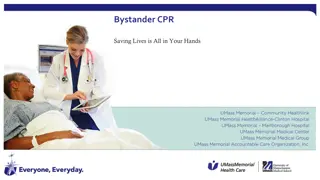
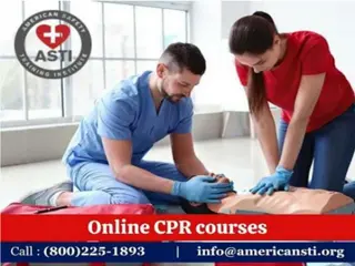
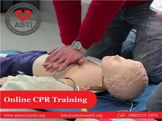
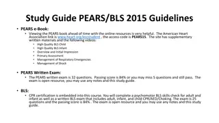
American Heart CPR & BLS: Emergency Response Steps
Learn the steps to activate the emergency response system in a cardiac emergency, including CPR procedures and AED usage. Follow the detailed instructions on assessing for breathing, giving breaths, performing compressions, and using an AED. Be prepared to act in situations where a person is unresponsive and not breathing.

Download Presentation

Please find below an Image/Link to download the presentation.
The content on the website is provided AS IS for your information and personal use only. It may not be sold, licensed, or shared on other websites without obtaining consent from the author.If you encounter any issues during the download, it is possible that the publisher has removed the file from their server.
You are allowed to download the files provided on this website for personal or commercial use, subject to the condition that they are used lawfully. All files are the property of their respective owners.
The content on the website is provided AS IS for your information and personal use only. It may not be sold, licensed, or shared on other websites without obtaining consent from the author.
E N D
Presentation Transcript
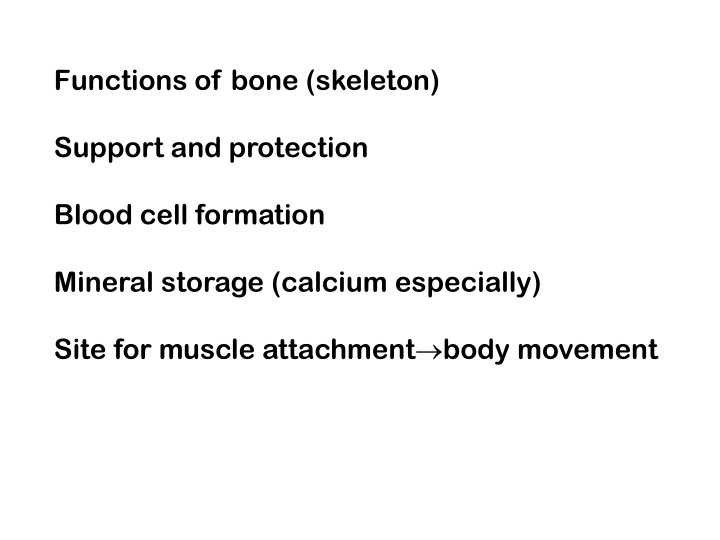
Functions of bone (skeleton) Support and protection Blood cell formation Mineral storage (calcium especially) Site for muscle attachment body movement
Bones classified by shape: long, short, flat, irregular, round Bone enclosed in periosteum, which is continuous with tendons and ligaments blood vessels in periosteum Epiphysis- ends spongy bone contains red marrow compact bone, articular cartilage Diaphysis- middle compact bone medullary cavity- contains yellow marrow (fat) lined with endosteum (squamous epithelium)
Compact bone osteocytes within lacunae arranged in concentric circles called lamellae This surround a central canal; complex is called Haversian system Canaliculi connect osteocytes to central canal and to each other
Prenatal development skeleton is mostly cartilaginous Cartilage cells and then osteoblasts start to deposit minerals Cartilaginous disk (epiphyseal disk) remains in epiphysis Cells eventually stop dividing
Adults continually break down and build up bone Osteoclasts remove damaged cells and release calcium into blood Osteoblasts remove calcium from blood and build new matrix. They become trapped osteocytes
Types of bone breaks Simple- skin is not pierced Compound- skin is pierced Complete- bone is broken in half Greenstick- incomplete break on outer Comminuted- broken into several pieces
Fracture repair Hematoma- blood clot in space between edges of break Fibrocartilage callus- begins tissue repair Bony callus- osteoblasts produce trabeculae (structural support) of spongy bone and replace fibrocartilage Remodeling- osteoblasts build new compact bone, osteoclasts build new medullary cavity
Axial skeleton skull (cranium and facial bones) hyoid bone (anchors tongue and muscles associated with swallowing) vertebral column (vertebrae and disks) thoracic cage (ribs and sternum) Appendicular skeleton pectoral girdle (clavicles and scapulae) upper limbs (arms) pelvic girdle (coxal bones, sacrum, coccyx) lower limbs (legs)
posterior view p. 135
Axial skeleton supports and protects organs of head, neck and trunk Appendicular skeleton- bones of limbs and bones that anchor them to the axial skeleton Articulation- where joints are formed
22 bones in skull 6 in middle ears 1 hyoid bone 26 in vertebral column 25 in thoracic cage 4 in pectoral girdle 60 in upper limbs 60 in lower limbs 2 in pelvic girdle 206 bones in all
The skull 8 sutured bones in cranium Facial bones: 13 bones, 1 mandible Cranium encases brain attachments for muscles sinuses
Vertebral column 7 cercvial vertebrae 12 thoracic 5 lumbar 1 sacrum (5 fused 1 coccyx (4 fused)
Thoracic cage ribs thoracic vertebrae sternum costal cartilages True ribs are directly attached to the sternum (first seven pairs) Three false ribs are joined to the 7th rib Two pairs of floating ribs
Clavicles and scapulae Help brace shoulders Attachment sites for muscles
Bones of upper limb Humerus (upper arm) Radius; ulna Carpals, metacarpals, phalanges Bones of lower limb Femur Patella Tibia, fibula Tarsals, metatarslas, phalanges
")