Anaemia in animals is characterized by a decrease in red blood cell mass caused by various factors like loss, destruction, or insufficient production. Learn about the classification, causes, and management of both regenerative and non-regenerative anaemia in veterinary medicine. Explore the developmental process of red blood cells and understand conditions leading to regenerative anaemia such as blood loss and hemolysis, and toxic causes of hemolysis. Delve into the details provided by Dr. Pallav Shekhar, Assistant Professor in Veterinary Medicine.
Please find below an Image/Link to download the presentation.
The content on the website is provided AS IS for your information and personal use only. It may not be sold, licensed, or shared on other websites without obtaining consent from the author. If you encounter any issues during the download, it is possible that the publisher has removed the file from their server.
You are allowed to download the files provided on this website for personal or commercial use, subject to the condition that they are used lawfully. All files are the property of their respective owners.
The content on the website is provided AS IS for your information and personal use only. It may not be sold, licensed, or shared on other websites without obtaining consent from the author.
E N D
Presentation Transcript
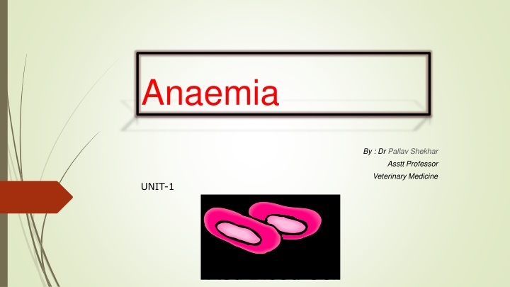
Anaemia By : Dr Pallav Shekhar Asstt Professor Veterinary Medicine UNIT-1
Definition and Etiology Anaemia is defined as an absolute decrease in the red cell mass as measured by RBC count, hemoglobin concentration, and PCV. It can develop from loss, destruction, or lack of production of RBC
Regenerative anaemia In a regenerative anemia, the bone marrow responds appropriately to the decreased red cell mass by increasing RBC production and releasing reticulocytes. Anaemias due to hemorrhage or hemolysis are usually regenerative.
Non regenerative anaemia In a non regenerative anaemia, the bone marrow responds inadequately to the increased need for RBC. Anaemias erythropoietin or an abnormality in the bone marrow are non regenerative that are caused by decreased
Non-regenerative anaemia Non- Regenerative anaemia Nutritional deficiencies Chronic diseases Renal diseases Chronic Inflammation, infection, neoplasia, liver disorder, Hypo- hyperadrenocorticism, hypothyroidism Fe, Cu, Vit B12, B6, Riboflavin, Niacin, Vit E & Vit C def. Chronic renal disease
Clinical signs : Lethargy and exercise intolerance Mucous membrane pallor Tachycardia Low grade haemic murmur Prominent femoral pulse Tachypnoea Episodic collapse (excitement or stress-induced ) Jaundice (in haemolytic anemia)
Pathogenesis of anemia Reduced oxygen carrying capacity of the blood. Inadequate tissue oxygenation Development of clinical signs Compensatory mechanism
Diagnosis: Hb and PCV value measurement Complete Blood Count Red Blood Cell morphology Reticulocyte count Erythrocytic Indices Platelet count
Red cell morphology Type of anemia Red cell morphology Acute blood loss Normocytic , normochromic No evidence of regeneration for 3-4 days. Chronic blood loss Hypochromasia, microcytic if iron deficiency present Hemolytic Spherocytosis Non-regenerative Normocytic, normochromic
Reticulocyte count Indicates degree of regeneration Erythrocytic Indices Mean Corpuscular Volume (MCV) Mean Corpuscular Hemoglobin Concentration (MCHC) Platelet count < 20,000/ l Thrombocytopenia DIC Intramedullary or extramedullary suppression of platelet production.
Faecal examination/ faecal occult blood test Severe upper gastrointestinal bleeding - black, tarry feces Bone marrow Evaluation Nonregenerative anemia Leukopenia Unexplained thrombocytopenia Pancytopenia
Treatment of anemia Emergency stabilization Blood transfusion Specific treatment of underlying cause
Blood transfusion PCV < 15% Selection of donor animals Major and minor cross Blood collection Blood transfusion
Other treatments- Anabolic Steroids Nandrolone decanoate ( 1 to 2 mg/kg/week, im Oxymetholone 1-5 mg/kg, PO, every 18-24 hr Stanozolol (dogs: 1-4 mg, PO, bid)
Hemostatics Astringents Epinephrine and norepinephrine Vitamin K Calcium therapy Desmopressin , Component therapy: Fresh Frozen Plasma and Platelet therapy


![Enhancing Patient Blood Management in Pregnancy at [INSERT LOCAL HOSPITAL NAME]](/thumb/143207/enhancing-patient-blood-management-in-pregnancy-at-insert-local-hospital-name.jpg)