Anatomy of Respiratory System: Features and Function
The respiratory system is vital for gas exchange between blood and air. Understanding the major anatomical features such as lung ventilation, gas exchange, and tissue interaction is crucial. Learn about how air is cleaned, distributed, and exchanged within the respiratory system. Explore how cilia clean the lungs, the role of alveoli in gas exchange, and the mechanics of ventilatory processes. Discover the organs involved in air movement and the significance of mucus in maintaining respiratory health.

Download Presentation

Please find below an Image/Link to download the presentation.
The content on the website is provided AS IS for your information and personal use only. It may not be sold, licensed, or shared on other websites without obtaining consent from the author.If you encounter any issues during the download, it is possible that the publisher has removed the file from their server.
You are allowed to download the files provided on this website for personal or commercial use, subject to the condition that they are used lawfully. All files are the property of their respective owners.
The content on the website is provided AS IS for your information and personal use only. It may not be sold, licensed, or shared on other websites without obtaining consent from the author.
E N D
Presentation Transcript
Lung and Respiration February 28 This lecture content will not be on Fridays test! What are the major anatomical features of the respiratory system. How is air cleaned prior to gas exchange? How is air distributed into smaller and smaller airways? What special structures in a trachea are related to the function of a trachea? How do cilia clean dirt and other materials from the lung? How does alveolar structure promote gas exchange? Remember: oxygen consumption is what it is all about! It all comes down to milliliters of O2/kg-minute Open Lab:: Turn Over a Better Leaf next week . MTWTh 6-8am and 6-8pm, Fri 6am-4pm
Anatomy and Physiology 212: Thinking and Learning go together for Test/Quiz scores A lot of the time we spend studying should be spend simply thinking try thinking about what you learned every night after lecture. On Average each Week (not a couple nights before), don t do it week before, study weekS before: 3 hours study time for each hour of lecture 3 X3 =9hours/wk 2 hours open lab for each hour in lab 2 X 2 = 4hours/wk in open lab Use of Quizlets, rewriting, conversations, etc. all help you to invest time in understanding lecture/lab content.
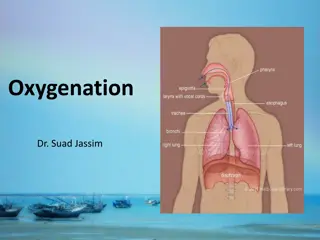
The Respiratory System provides for gas exchange between the blood and air. Respiration has three different connotations: 1) Lung Ventilation: 2) Lung Gas Exchange: 3) Tissue: oxygen carbon dioxide and energy Upper Respiratory Tract: Head and Neck Filtering out harmful particles in air Humidifying air Delivering air to the lung Lower Respiratory Tract: Thorax Gas Exchange Prevention of infection Clean alveolar surface Ventilatory Mechanics: Skeletal muscle of diaphragm and intercostal muscles between ribs. Also: abdomenal muscles Most people only use energy to inhale ! (Exhale for free)
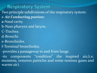
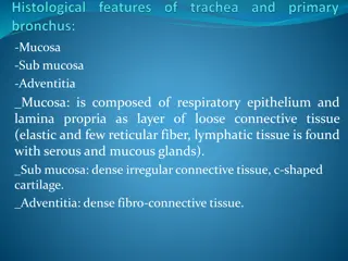
What organs facilitate the movement of air to the lung? The importance of mucus and the mucus membrane: CRITICAL! Air Entry: Nasal Cavity and Conchae- Sinus Cavities- Oral Cavity: poor airflow efficiency/low resistance Pharynx- Epiglottis and Glottis- Trachea- Bronchial Tree- Alveolus- Each lung and its lobes sit in a pleural cavity under a slight vacuum Esophogus is often mistaken for an airway during anaesthesia, this can be fatal!
Overview of the primary airway structures. Each lung (Rt/Lt) is surrounded by pleural fluids and sits in a pleural cavity that is under a slight vacuum. V.I.P: Pleural Cavity under vacuum
Air is distributed into lower respiratory air passages called bronchi and bronchioles that ultimately lead to dead-end alveoli. The Bronchial Tree : moves air from large to narrow branching airways Bronchi with cartilage for support and smooth muscle: Primary B Secondary B Tertiary B Bronchioles Have cilia for cleaning purposes Nourished by bronchiolar artery Bronchioles are 1mm diameter airways with smooth muscle for changing diameter to needs Bronchiole TerminalBronchiole RespiratoryBronchiole Alveoli(Gas Exchange) Respiratory Bronchiole and alveoli have no cilia or smooth muscle! The smooth muscle lining Bronchiole can constrict Asthma Alveoli: clusters of dead end pouches for gas exchange! Alveolar macrophages: cleaning Type II alveolar cells: surfactant Type I alveolar cells (simple squamous epithelial): gas exchange
Bronchiole Terminal Bronchiole Respiratory Bronchiole Alveolar Sacs
Amazing Statistics about the lung: A fully inflated lung can contain about 5-6liters of air. The surface area of the 500 million tiny alveoli in the two lungs is about 75 meters2 The surface area of the capillaries that sit underneath the alveoli is about 60 meters2 for a single lung. Surprisingly the volume of blood in these alveolar capillaries at rest is only about 70 ml and perhaps 200 ml when exercising rigorously. Clearly the blood must move through these capillaries very rapidly and gas exchange must ALSO be very rapid (transit time across the alveoli is 0.75 seconds or less).
The shape of the lung describes why we sometimes have respiratory difficulties. Each lung sits in a pleural cavity, you have two pleural cavities in your thorax. Why do right and left lung lobes have different sizes? Importance of having 2 lobes sealed separately? Pneumothorax: if pleural cavity loses its vacuum, the lung collapses and gas exchange to blood stops! Implications: Cancer? Infection? GunShot (deer/human)? Why do aspirated objects/obstructions affect the right side more often than the left? A story about kids eating marbles! If you have to have a lung removed, why does left lung removal affect you less than right lung removal?
The respiratory system is rich in elastin and cartilage for two very different reasons. Why is elastin so important? It stretches and stretches back! Located in lung tissue and thoracic wall! Inhalation: Active Exhalation: Passive Emphysema/cigarette smoke/ alpha-1-antitrypsin/elastin loses Barrel Chest: sign of loss of elastin Why is cartilage so important? Hyalin Rings: trachea and bronchi of lung Cartilagenous rings prop airways open and keep airways from collapsing when you inhale Smooth muscle in airways lets you control tune diameter/airflow Cartilaginous hyalin rings help prevent airway collapse when you INSPIRE! Why? Elastin causes the lung to collapse passively (exhale)! WHY?
Asthma: trachealis and other airway smooth muscle cells constrict in response to irritants, this increases airway resistance. Q r4 V.I.P Changes in resistance are most pronounced when the bronchioles constrict. Asthma can also be protective sometimes .why?
Cilia are tiny hair-like structures that carry mucus, bacteria, pollen, dust and other potentially harmful material (caught in mucus) up and out to the glottis. However, cilia do not extend into the alveolus/terminal bronchiole and can be destroyed by smoke! Why do you sometimes have to cough ? Why do smokers cough more often?
Lungs move and circulate air to maximize alveolar gas exchange. Unfortunately we are not efficient at doing this. Primary, Secondary and Tertiary Bronchi -Cilia, SMC, non-respiratory and brachial artery -Acute effects of asthma found here! Terminal Bronchioles and Respiratory Bronchioles ------Scant-gas exchange/ no or few cilia /No or few SMCs Until this point the air sits in what can be called a dead space (called dead because significant gas exchange can t occur here). These dead spaces should never collapse. You CAN inhale air and get no gas exchange to the blood because of collapsed bronchioles due to asthma!
Terminal and respiratory bronchioles have very short lengths that terminate in the blind alveolar sacs. Each sac is richly supplied with a capillary bed for gas transfer to/from blood.
In our Respiratory Physiology lab we will use an electronic spirometer that converts air velocity and flow into respiratory volumes using a set of computer algorithms. Old School Spirometer