Anatomy of the Tongue: Muscles, Mucous Membrane, and Blood Supply
Explore the intricate anatomy of the tongue, including details on its muscular structure, mucous membrane composition, and blood supply. Learn about the division of the tongue into anterior and posterior parts, the presence of papillae, and the role of intrinsic and extrinsic muscles. Discover how the lingual artery, facial artery, pharyngeal artery, and internal jugular vein contribute to the blood supply of the tongue.
Download Presentation

Please find below an Image/Link to download the presentation.
The content on the website is provided AS IS for your information and personal use only. It may not be sold, licensed, or shared on other websites without obtaining consent from the author. If you encounter any issues during the download, it is possible that the publisher has removed the file from their server.
You are allowed to download the files provided on this website for personal or commercial use, subject to the condition that they are used lawfully. All files are the property of their respective owners.
The content on the website is provided AS IS for your information and personal use only. It may not be sold, licensed, or shared on other websites without obtaining consent from the author.
E N D
Presentation Transcript
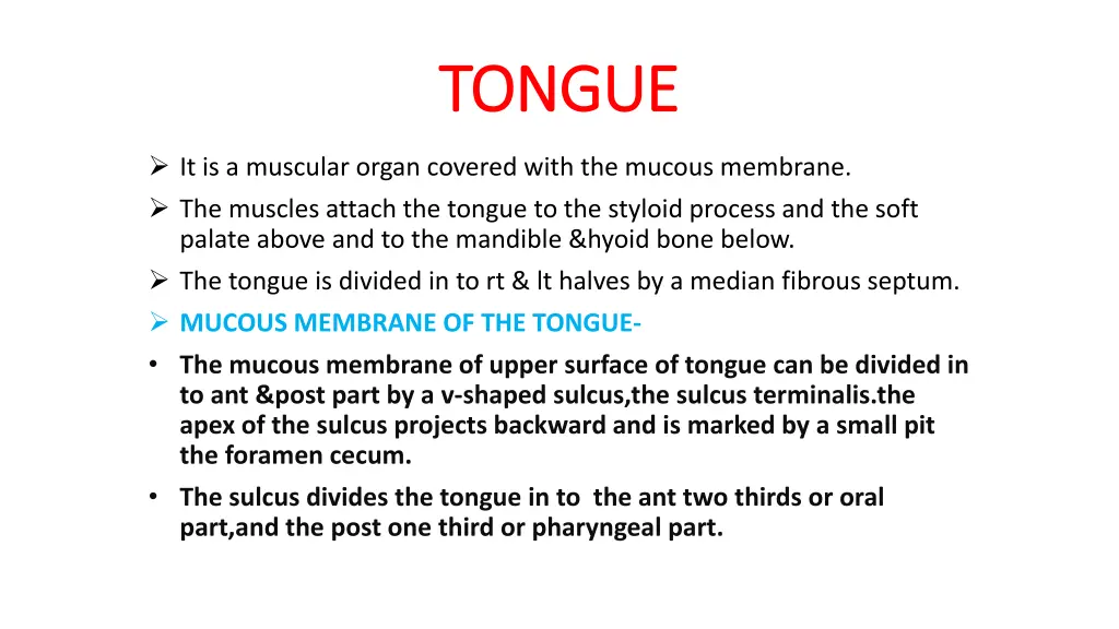
TONGUE TONGUE It is a muscular organ covered with the mucous membrane. The muscles attach the tongue to the styloid process and the soft palate above and to the mandible &hyoid bone below. The tongue is divided in to rt & lt halves by a median fibrous septum. MUCOUS MEMBRANE OF THE TONGUE- The mucous membrane of upper surface of tongue can be divided in to ant &post part by a v-shaped sulcus,the sulcus terminalis.the apex of the sulcus projects backward and is marked by a small pit the foramen cecum. The sulcus divides the tongue in to the ant two thirds or oral part,and the post one third or pharyngeal part.
MUCOUS MEMBRANE CONT MUCOUS MEMBRANE CONT- - The foramen cecum is an embryonic is an embryonic remnant and marks the site of upper end of the thyroglossal duct. Three types of papillae are present on the upper surface of the ant two thrds of the tongue: the filiform papillae,the fungiform papillae,and the vallate papillae. The mucous membrane covering the post third of the tongue is devoid of papillae but has an irregular surface, caused by the presence of underlying lymph nodules,the lingual tonsil.
The mucous membrane on the inferior surface of the tongue is reflected from the tongue to the floor of the mouth. In the mid line anteriorly,the undersurface of the tongue is connected to the floor of the mouth by a fold of mucous membrane,the frenulum of the tongue. On the lateral side of the frenulum,the deep lingual vein can be seen through the mucous membrane. Lateral to the lingual vein,the mucous membrane forms a fringe fold called the plica fimbriata.
MUSCLES OF THE TONGUE MUSCLES OF THE TONGUE The muscles of the tongue are divided in to two types: intrinsic and extrinsic. INTRINSIC MUSCLES- These muscles are confined to the tongue and are not attached to bone. They consist of longitudinal,transverse and vertical fibres. Nerve supply-hypoglossal nerve. Action-alter the shape of the tongue.
EXTRINSIC MUSCLES EXTRINSIC MUSCLES These muscles are attached to bones and the soft palate. They are the genioglossus,the hyoglossus,the styloglossus and the palatoglossus NERVE SUPPLY-All the muscles are supplied by hypoglossal nerve except palatoglossus which is supplied by pharyngeal plexus. BLOOD SUPPLY-The lingual artery,the tonsillar branch of the facial artery,and the ascending pharyngeal artery supply the tongue.The veins drain in to the internal jugular vein.
BLOOD SUPPLY BLOOD SUPPLY BLOOD SUPPLY- The lingual artery, The tonsillar branch of the facial artery,and The ascending pharyngeal artery supply the tongue. The veins drain in to the internal jugular vein.
LYMPHATIC DRAINAGE TIP-submental lymph nodes. sides of the ant two thirds-submandibular and deepcervical lymph nodes. posterior third: deep cervical lymph nodes. SENSORY INNERVATION- ANTERIOR TWO THIRDS-Lingual nerve branch of mandibular division of trigeminal nerve(general sensation)&chorda tympani branch of the facial nerve(taste). POSTERIOR THIRD-glossopharyngeal nerve(general sensation & taste)
MOVEMENTS OF THE TONGUE PROTRUSION-the genioglossus muscles on both sides acting together. RETRACTION-styloglossus &hyoglossus muscles on both sides acting together. DEPRESSION-hyoglossus muscles on both sides acting together. ELEVATION-styloglossus and palatoglossus muscles on both sides acting together.
DEVELOPMENT OF TONGUE DEVELOPMENT OF TONGUE Tongue muscles are derived from occipital myotomes which migrates forward carrying their nerve supply with them(hypoglossal nerve). The epithelium is derived from the lining of the floor of the pharynx and comes from parts of first,third and fourth arches. The mucosa of the ant two thirds is from the ectodermal lining of the midline tuburculum impar and the pair of lateral lingual swellings of the first arch(lingual and chorda tympani nerve). The mucosa of posterior third is derived from the endodermal lining of the midline hypobranchial eminence of the third arch(glossopharyngeal nerve),with a small contribution from the fourth arch(internal laryngeal nerve). Tissue of the second arch is not represented because third arch tissue over grows it in a forward direction to meet that from the first arch.


