Anatomy of Trachea: Structure and Relations
The trachea, also known as the air vessel, is a vital structure in the neck and mediastinum. Learn about its dimensions, relations, and course in the body. Explore its role in breathing and the anatomical features that make it unique.

Download Presentation

Please find below an Image/Link to download the presentation.
The content on the website is provided AS IS for your information and personal use only. It may not be sold, licensed, or shared on other websites without obtaining consent from the author.If you encounter any issues during the download, it is possible that the publisher has removed the file from their server.
You are allowed to download the files provided on this website for personal or commercial use, subject to the condition that they are used lawfully. All files are the property of their respective owners.
The content on the website is provided AS IS for your information and personal use only. It may not be sold, licensed, or shared on other websites without obtaining consent from the author.
E N D
Presentation Transcript
ANATOMY OF TRACHEA By : Dr. Manjula Vastrad Asst Prof Dept of Rachana Shareera SMVVS RKM AMC VIJAYAPUR
INTRODUCTION Trachea (In latin air vessel) is a wide tube Lying more or less in midline, in the lower part of neck and in the superior mediastinum
Its upper end is continuous with with the lower end of larynyx Trachea in the neck is covered by isthmus of the thyroid gland and acts as a sheild for trachea At its lower end the trachea ends by dividing into right and left principal bronchi
DIMENSIONS Trachea is 10 15 cm in length Its external diameter measures about 2cm in males and 1.5cm in females The lumen is smaller in living than in cadever Its about 3mm at one year of age During childhood it corresponds to the age in years, with the max of about 12mm in adults i.e it increases 1mm per year upto 12 years
RELATIONS The upper end of trachea lies at the lower border of cricoid cartilage, opp C6 vertebra In the cadever its bifurcated lower end lies at the lower border of T4 vertebra, corresponding in front to sternal angle However in living subjects, in the erect posture, the bifurcation lies at the lower border of T6 vertebra and descends still further during inspiration
COURSE Over most of its length trachea lies in median plane But near the lower end it deviates slightly to the right As it runs downwards, the trachea passes slightly backwards following the curvature of spine
RELATIONS IN THORAX Anteriorly Manubrium sterni Sternohyoid and Sternothyroid muscles Remains of thymus Left brachiocephalic and inferior thyroid veins Aortic arch, brachiocephalic and left common carotid arteries Some lymph nodes
Posteriorly Oesophagus Vertebral column Right side Right lung and pleura Right vagus Azygous vein Left side Arch of aorta, left common carotid and left subclavian arteries Left recurrent laryngeal nerve
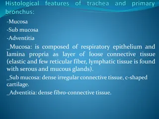
STRUCTURE Trachea has fibroelastic wall supported by a cartilagenous skeleton formed by C-shaped rings Rings are about 16-20 in number and make the tube convex anterolaterally Posteriorly there is a gap which is closed by fibroelastic membrane and contains transversely arranged smooth muscle known as trachealis The lumen is lined by ciliated columnar epithelium and contains many mucous and serous glands
Widest ring 1string Carina a hook shaped triangular process upwards from the lower part of last ring at the bifurcation and this ring surrounds the two bronchi It presents a ridge in the interior of trachial bifurcation. This ridge is a guide to surgeon during bronchoscopic and other examination Functions Cartilages prevent collapsing of trachea and so keep the trachea patent Posteriorly cartilages are deficient for allowing expansion of oesophagus during swallowing
ARTERIAL SUPPLY Inf thyroid artery branch, of thyrocervical trunk Bronchial arteries (at the bifurcation of trachea) VENOUS DRAINAGE Into inf thyroid venous plexus
LYMPHATICS Pretracheal and paratracheal lymph nodes NERVE SUPPLY Parasympathetic From rt and left vagus nerves Recurrent laryngeal nerves Sympathetic From upper 4-5 thoracic segments of spinal cords
APPLIED ANATOMY Tracheoesophageal fistula Congenital anamoly where communication occur b/w the bifurcation of trachea and oesophagus
Carina present in the last ring of trachea helps in bronchoscopic examination Foreign body in trachea Any foreign body through trachea will enters into the right bronchus more easily, as it is more vertical
The posterior part is flattened which helps the passage of food bolus through the oesophagus Tracheostomy Is done by the midline incision with isthmus of thyroid gland retracted inferiorly Indications In adult: severe laryngeal damage In infant : severe airway obstruction Site Through 2ndtracheal ring then tracheostomy tube is inserted
Trachea may get compressed by pathological enlargements of the thyroid , thymus, lymph nodes and the aortic arch. This causes dyspnoea, irritative cough, and often a husky voice
THANK YOU