Anesthesia Techniques Systemic Circulation Overview
Systemic circulation is a vital part of the cardiovascular system, responsible for transporting oxygenated blood away from the heart to the body and returning deoxygenated blood back. It includes arteries, arterioles, capillaries, venules, and veins, each playing a crucial role in this process. Arteries carry oxygenated blood from the heart through the aorta, while arterioles control blood flow into capillaries. Capillaries are thin vessels that facilitate nutrient transport to tissues, and venules collect deoxygenated blood before it reaches the heart through veins. Understanding the systemic circulation is fundamental for grasping cardiovascular physiology.
Download Presentation

Please find below an Image/Link to download the presentation.
The content on the website is provided AS IS for your information and personal use only. It may not be sold, licensed, or shared on other websites without obtaining consent from the author. If you encounter any issues during the download, it is possible that the publisher has removed the file from their server.
You are allowed to download the files provided on this website for personal or commercial use, subject to the condition that they are used lawfully. All files are the property of their respective owners.
The content on the website is provided AS IS for your information and personal use only. It may not be sold, licensed, or shared on other websites without obtaining consent from the author.
E N D
Presentation Transcript
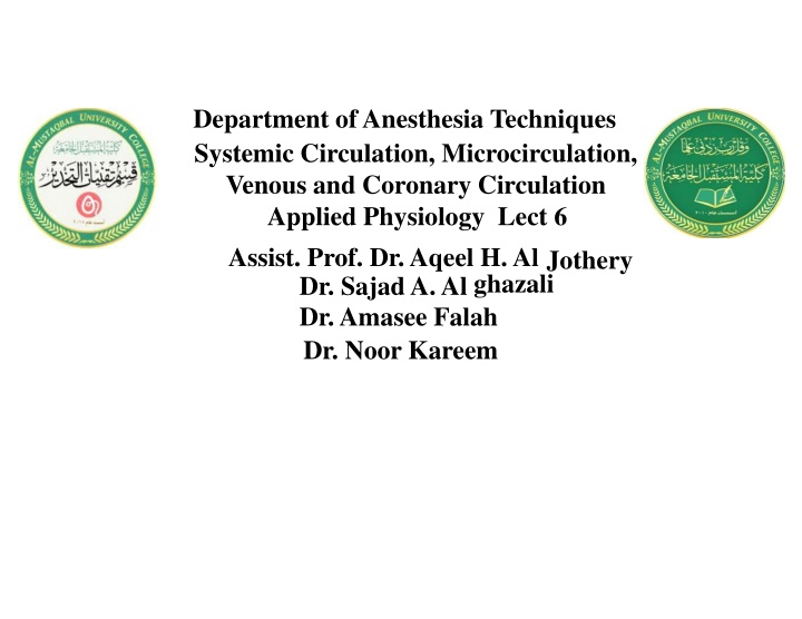
Department of Anesthesia Techniques Systemic Circulation, Microcirculation, Venous and Coronary Circulation Applied Physiology Lect 6 Assist. Prof. Dr. Aqeel H. AlJothery Dr. Sajad A. Alghazali Dr. Amasee Falah Dr. Noor Kareem
Systemic circulation :is the portion of the cardiovascular system which carries oxygenated blood away from the heart, to the body, and returns deoxygenated blood back to the heart. The term is contrasted with pulmonary circulation. Systemic circulation include 5 types of blood vessels a. Arteries b. Arterioles c. Capillaries d. Venules e. Veins
A-Arteries Oxygenated blood enters the systemic circulation when leaving the left ventricle, through the aortic semi-lunar valve. The first part of the systemic circulation is the artery aorta, a massive and thick-walled artery. The aorta arches and gives off major arteries to the upper body before piercing the diaphragm in order to supply the lower parts of the body with its various branches. B-Arterioles The smallest branches of the arteries. They role consist in controlling the flow of blood into the capillary system. This is achieved by an action of muscle cells in their walls. Arterioles and small arteries are known as resistance vessels, because they form a main portion of a peripheral resistance value.
C-Capillaries Blood passes from arteries to capillaries, which are the thinnest and most numerous of the blood vessels. These capillaries help to join tissue with arterioles for transportation of nutrition to the cells, which absorb oxygen and nutrients in the blood. Peripheral tissues do not fully deoxygenate the blood, so venous blood does have oxygen, but in a lower concentration than in arterial blood. In addition, carbon dioxide and wastes are added. D-Venules The deoxygenated blood is then collected by venules, from where it flows first into veins, and then into the inferior and superior venae cavae, which return it to the right heart, completing the systemic cycle. The blood is then re-oxygenated through the pulmonary circulation before returning again to the systemic circulation.
E-Veins The relatively de-oxygenated blood collects in the venous system which coalesces into two major veins: the superior vena cava (roughly speaking from areas above the heart) and the inferior vena cava (roughly speaking from areas below the heart). These two great vessels exit the systemic circulation by emptying into the right atrium of the heart. The coronary sinus empties the heart's veins themselves into the right atrium.
Microcirculation and Starling law of the capillaries The microcirculation is responsible for the transfer of oxygen from the red blood cells in the capillaries to the cells to meet cellular energy requirements, support functional activity and remove carbon dioxide and waste. The microcirculation also assists in the regulation of vascular tone, solute exchange, the production of hormones, the inflammatory response and haemostasis Elements of Microcirculation Arterioles, capillaries, venules Structure and function: transport Fluid balance
The principal function of the microcirculation is to permit the transfer of substances between the tissues and the circulation. This transfer occurs predominantly across the walls of the capillaries but some exchange occurs in the small venules also. Substances involved include water, electrolytes, gases (O2, CO2), nitrogenous wastes, glucose, lipids and drugs. Exchange of Water, Nutrients, and Other Substances Between the Blood and Interstitial Fluid Achieved By two processes 1-Diffusion through Capillaries Dominant mechanism, rate varies by tissue Diffusion of water 40-80 times faster than flow 2/3 of blood volume diffuses per minute Driven by concentration gradient: J = -DAdc/dx D = diffusion constant,A= area, c = concentration Flow limited: transport rate is fast enough for equilibrium Diffusion limited: equilibrium never established LipidSoluble O2 and CO2 H2O H2O-soluble small H2O-soluble medium H2O-soluble large
-Lipid-Soluble Substances Can Diffuse Directly Through the Cell Membranes of the Capillary Endothelium such as CO2 and O2 -Water-Soluble, Non-Lipid-Soluble Substances Diffuse Only Through Intercellular Pores in the Capillary Membrane such as sodium ions and glucose 2-Filtration through Capillaries: driven by Hydrostatic and Osmotic Pressure Fluid Balance: Hydrostatic Pressure Capillary Pressure (Pc) Interstitial Fluid Pressure (Pi) Hydrostatic pressure in the capillaries tends to force fluid and its dissolved substances through the capillary pores into the interstitial spaces
Fluid Balance: Osmotic Pressure Capillary Pressure (Pc) Plasma colloid osmotic Pressure ( c) Interstitial Fluid Pressure (Pi) Interstitial colloid osmotic Pressure ( i) Osmotic pressure caused by the plasma proteins (called colloid osmotic pressure) tends to cause fluid movement by osmosis from the interstitial spaces into the blood. Starling Law mentioned by a British Physiologist Starling (1896) states that the fluid movement due to filtration across the wall of a capillary is dependent on the balance between the hydrostatic pressure gradient and the oncotic pressure gradient across the capillary. The four Starling's forces are: hydrostatic pressure in the capillary (Pc), hydrostatic pressure in the interstitium (Pi), oncotic pressure in the capillary (pc ), oncotic pressure in the interstitium (pi)
hydrostatic pressure in the capillary (Pc)-tends to force fluid outward through the capillary membrane. hydrostatic pressure in the interstitium (Pi)-tends to force fluid inward through the capillary membrane oncotic pressure in the capillary (pc )-tends to cause osmosis of fluid inward through the capillary membrane. oncotic pressure in the interstitium (pi)-tends to cause osmosis of fluid outward through the capillary membrane. If the sum of these forces, the net filtration pressure, is positive, there will be a net fluid filtration across the capillaries. If the sum of the Starling forces is negative, there will be a net fluid absorption from the interstitial spaces into the capillaries.
The net filtration pressure (NFP) is calculated as: NFP= Pc-Pi + pc-pi the NFP is slightly positive under normal conditions, resulting in a net filtration of fluid across the capillaries into the interstitial space in most organs. The rate of fluid filtration in a tissue is also determined by the number and size of the pores in each capillary as well as the number of capillaries in which blood is flowing. These factors are usually expressed together as the capillary filtration coefficient (Kf). The Kf is therefore a measure of the capacity of the capillary membranes to filter water for a given NFP and is usually expressed as ml/min per mm Hg net filtration pressure. The rate of capillary fluid filtration is therefore determined as: Filtration= Kf x NFP
Venous circulation Functions of Veins: 1-Drainage of Blood from all parts of the body to the heart. 2-It act as blood reservoirs- contain about 3 litters of blood 3-Venous pump- aids in propelling the blood forward (towards the heart ) and helps to regulate the COP. Venous pressure It is the pressure inside veins (10-11 mmHg) a)Hydrostatic indifferent level or point (HIP) It lies 5-7 cm below the diaphragm, at which the VP is kept constant , independent of the body posture VP below this point high High , above this point Low b)Perpherial venous pressure (PVP) It depends upon the site of vein and the gravity -above right atrium VP is subatomsphric c-)Central venous pressure (CVP) It is VP in big veins at right atrium It an averages 4-6 mmHg in recumbancy and 2 mmHg on standing It is 2 mmHg during inspiration and 6 mmHg during expiration
Factors affecting venous return Right atrial pressure Total blood volume Venoconstriction Respiratory pump skeletal Muscle pump Gravity 14
1.Right atrial pressure: An increase in right atrial pressure decreases venous return. Venous return=arterial pressure- right atrial pr./Total peripheral resistance 2. Total blood volume : An increase in total blood volume increases venous return. B.V can be increased by I.V. fluid & blood transfusion 3. Venoconstriction: Constriction of the veins reduces the size of the venous reservoirs, decreasing venous pooling and thus increasing venous return 4. Respiratory pump: During inspiration, the venous return increases because of a decrease in right atrial pressure by the increased negative intra pleural pr. 5. skeletal Muscle pump: muscular activity increases venous return as a result of the pumping action of skeletal muscle. 6. Gravity: Standing decreases venous return
The Coronary Circulation The coronary circulation refers to the vessels that supply and drain the heart. Coronary arteries are named as such due to the way they encircle the heart, much like a crown. An arterial circle surrounds the heart in coronary (AV) sulcus. From this arterial circle an arterial loop runs in the anterior & inferior interventricular grooves.
The Arterial Supply of the Heart The Arterial Supply of the Heart is mostly supplied by 2 coronary arteries, which originate from the ascending aorta immediately above the aortic valve from the aortic sinuses. The coronary arteries and their branches run on the surface of heart being located inside the subpericardial fibrofatty tissue. Coronary arteries are vasa vasorum of the ascending aorta. Anatomically coronary arteries are not end-arteries but functionally they behave like end-arteries.Coronary Arteries There are two main coronary arteries which branch to supply the entire heart. They are named the left and right coronary arteries, and arise from the left and right aortic sinuses within the aorta. The aortic sinuses are small openings found within the aorta behind the left and right flaps of the aortic valve. When the heart is relaxed, the back-flow of blood fills these valve pockets, therefore allowing blood to enter the coronary arteries.
Characteristics of Coronary Arteries A. Coronary arteries are vasa vasorum of the ascending aorta. B. Anatomically coronary arteries are not end-arteries but functionally they behave like end-arteries. C. The only branches of ascending aorta. D. The only arteries which get filled up during diastole of heart.
Thank you for your attention

