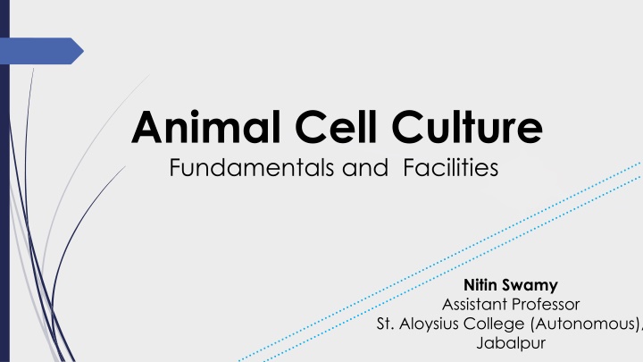
Animal Cell Culture Fundamentals and Facilities
Explore the fundamentals of animal cell culture, including its historical background, terminology, primary culture, and the importance of sub-culturing. Learn about the facilities required for successful animal cell culture in the laboratory setting.

Download Presentation

Please find below an Image/Link to download the presentation.
The content on the website is provided AS IS for your information and personal use only. It may not be sold, licensed, or shared on other websites without obtaining consent from the author. If you encounter any issues during the download, it is possible that the publisher has removed the file from their server.
You are allowed to download the files provided on this website for personal or commercial use, subject to the condition that they are used lawfully. All files are the property of their respective owners.
The content on the website is provided AS IS for your information and personal use only. It may not be sold, licensed, or shared on other websites without obtaining consent from the author.
E N D
Presentation Transcript
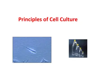
Animal Cell Culture Fundamentals and Facilities Nitin Swamy Assistant Professor St. Aloysius College (Autonomous), Jabalpur
Introduction to Animal Cell Culture Animal cell culture basically involves the in vitro (in the laboratory) maintenance and propagation of animal cells in a suitable nutrient media. Thus, culturing is a process of growing cells artificially. Historical Background It was in 1907, Ross Harrison first developed a frog tissue culture technique. In 1940 s chick embryo tissue became a favorite for culture techniques. Interest in culturing human tissues started in 1950 s after it was demonstrated (by HeLa; Gey) that human tumor cells could give rise to continuous cell lines. Among the various animal cell cultures, mouse cell cultures are the most commonly used in the laboratory.
Terminology in Cell Culture Organ culture The culture of native tissue (i.e. un-disaggregated tissue) that retains most of the in vivo histological features is regarded as organ culture. : Cell culture This refers to the culture of dispersed (or disaggregated) cells obtained from the original tissue, or from a cell line. : Histotypic culture The culturing of the cells for their re-aggregation to form a tissue like structure represents histotypic culture. : Organotypic culture This culture technique involves the recombination of different cell types to form a more defined tissue or an organ.
Primary culture: The culture produced by the freshly isolated cells or tissues taken from an organism is the primary culture. These cell are heterogenous and slow growing, and represent the tissue of their origin with regard to their properties. Cell line: The sub-culturing of the primary culture gives rise to cell lines. The term continuous cell lines implies the indefinite growth of the cells in the subsequent sub-culturing. On the other hand, finite cell lines represent the death of cells after several subcultures.
Why sub culturing.? Once the available substrate surface is covered by cells (a confluent culture) growth slows & ceases. Cells to be kept in healthy & in growing state have to be sub- cultured or passaged It s the passage of cells when they reach to 80-90% confluency in flask/dishes/plates Enzyme such as trypsin, dipase, collagenase in combination with EDTA breaks the cellular glue that attached the cells to the surface
Facilities for Animal Cell Culture While designing the laboratory for animal cell culture technology, utmost care should be taken with regard to the maintenance of aseptic conditions. The facilities required with regard to infrastructure and equipment are listed below : Minimal Requirements for Cell Culture i. Clean and quite sterile area ii. Preparation facilities iii. Animal house iv. Microbiology laboratory v. Storage facilities (for glassware, chemicals, liquids, small equipment). Equipment Laminar-flow, sterilizer, incubator, refrigerator and freezer (-20 C), balance, C02 cylinder, centrifuge, inverted hemocytometer, liquid nitrogen freezer, slow- cooling device (for freezing cells), pipette washer, deep washing sink. Besides the basic and minimal requirements listed above, there are many more facilities that may be beneficial or useful for tissue cultures. These include air-conditioned rooms, containment room for biohazard work, phase-contrast microscope, fluorescence microscope, confocal microscope, and high capacity centrifuge and time lapse video equipment. microscope, water purifier,
Culture Vessels: In the tissue culture technology, the cells attach to the surface of a vessel which serves as the substrate, and grow. Hence there is a lot of importance attached to the nature of the materials used and the quality of the culture vessels. The term anchorage dependent cells is used when the cells require an attachment for their growth. On the other hand, some cells undergo transformation, and become anchorage independent. Materials used for culture vessels Glass: Although glass was the original substrate used for culturing, its use is almost discontinued now. This is mainly because of the availability of more suitable and alternate substrates. Disposable plastics: Synthetic plastic materials with good consistency and optical properties are now in use to provide uniform and reproducible cultures. The most commonly used plastics are polystyrene, polyvinyl chloride (PVC), polycarbonate, metinex and thermonex (TPX). Types of culture vessels The following are the common types of culture vessels. i. Multiwall plates ii. Petridishes iii. Flasks iv. Stirrer bottles.
Advantages of Tissue Culture The most important advantages of this technique are listed below 1. Control of physicochemical environment- pH, temperature, dissolved gases (O2 and CO2), osmolarity. 2. Regulation of physiological conditions-nutrient concentration, cell to cell interactions, hormonal control. 3. The cultured cell lines become homogenous (i.e. cells are identical) after one or two subcultures. This is in contrast to the heterogenous cells of tissue samples. The homogenous cells are highly useful for a wide range of purposes. 4. It is easy to characterize cells for cytological and immunological studies.
5. Cultured cells can be stored in liquid nitrogen for several years. 6. Due to direct access and contact to the cells, biological studies can be carried out more conveniently.The main advantage is the low quantities of the reagents required in contrast to in vivo studies where most of the reagents (more than 90% in some cases) are lost by distribution to various tissues, and excretion. 7. Utility of tissue cultures will drastically reduce the use of animals for various experiments.
Limitations of Tissue Culture There are several limitations of tissue culture; some of them are given below. 1. Need of expertise and technical skill for the development, and regular use of tissue culture. 2. Cost factor is a major limitation. Establishment of infrastructure, equipment and other facilities are expensive. 3. It is estimated that the cost of production of cells is about 10 times higher than direct use of animal tissues. 4. Control of the environmental factors (pH, temperature, dissolved gases, disposal of biohazards) is not easy. 5. The native in vivo cells exist in a three- dimensional geometry while in in vitro tissue culture, the propagation of cells occurs on a two dimensional substrate. Due to this, the cell to cell interactive characters are lost.
6. The cell lines may represent one or two types of cells from native tissue while others may go unrepresented. the 7. Tissue culture techniques are associated with the differentiation i.e. loss of the characters of the tissue cells from which they were originally isolated. 8. This happens due to adaptation and selection processes while culturing. 9. Continuous cell lines may result in genetic instability of the cells. This may ultimately lead to heterogeneity of cells. 10. The components of homeostatic in vivo regulation (nervous system, endocrine system, lacking in vitro cultures. Addition of hormones and growth factors has been started recently. metabolic integration) are
Mamalian cells are divided by Normal (mortal) and immortal (continuous/transformed) Normal: Divide only for limited of generation (30generations). Transformed: Can be propagated
Major developments in cell culture technology First development was the use of antibiotics which inhibits the growth of contaminants. Second was the use of trypsin to remove adherent cells to subculture further from the culture vessel Third was the use of chemically defined culture medium.
A growth medium or culture medium is a liquid or gel designed to support the growth of cells Types of Cell Culture Media Media Type Examples plasma, serum, lymph, human placental cord serum, amniotic fluid Biological Fluids Extract of liver, spleen, tumors, leucocytes and bone marrow, extract of bovine embryo and chick embryo Natural media Tissue Extracts Clots coagulants or plasma clots Balanced salt solutions PBS, DPBS, HBSS, EBSS Basal media MEM DMEM Artificial media Complex media RPMI-1640, IMDM
Natural Media Very useful Lack of knowledge of the exact composition of these natural media In this type of media used tissue extract, like liver, spleen, bone marrow, chick embryo extract and other body fluids like amniotic fluid and lymph fluid etc.
Artificial Media Serum containing media Serum-free media (defined culture media) Chemically defined media Protein-free media Artificial media/ defined media basic benefit of this media is that has all define nutrients but pH of this should be approx 7. Purpose of this type media using that provided all known nutrients to cells we also can include some other nutrients which cause cell proliferation and differentiation.
Serum free media- in this media using the serum as nutrient base for cells .benefits of this we can control to cell growth and also include cell proliferation factor and differentiation factors in it. But this has also disadvantage like slow growth rate, and require reagents totally pure conditions. Serum media- Is the example of natural media . Role of this in cell culture is very complex that also has some growth inhibitor which affect to cell culture. serum help to cell attachment and work as spreading factor in culture. This work add in culture albumin carrier factor, that help in reduced to mechanical damage and also work as natural buffering agent, also help to maintain pH of media.
Basic Components of Culture Media Culture media (as a powder or as a liquid) contains: amino acids Glucose Salts Vitamins Other nutrients The requirements for these components vary among cell lines, and these differences are partly responsible for the extensive number of medium formulations .
Natural buffering system HEPES Phenol red as a pH indicator (yellow or purple) Inorganic salt Amino Acids (L-glutamine) Carbohydrates Proteins and Peptides (important in serum-free media. Serum is a rich source of proteins and includes albumin, transferrin, aprotinin, fetuin, and fibronectin Fatty Acids and Lipids Vitamins Trace Elements
Common Cell Culture Media Eagle s Minimum Essential Medium (EMEM) Dulbecco s Modified Eagle s Medium (DMEM) Low glucose High glucose RPMI-1640 Ham s Nutrient Mixtures DMEM/F12 Iscove s Modified Dulbecco s Medium (IMDM)
Cell Line Morphology Species Medium Applications MEM+ 2mM Glutamine+ 10% FBS + 1% Non Essential Amino Acids (NEAA) RPMI 1640 + 2mM Glutamine + 10- 20% FBS DMEM + 2mM Glutamine +5% New Born Calf Serum (NBCS) + 5% FBS Tumourigenicity and virus studies HeLa B Epithelial Human HL60 Lymphoblast Human Differentiation studies 3T3 clone A31 Tumourigenicity and virus studies Fibroblast Mouse Gene expression and virus replication studies COS-7 Fibroblast Monkey DMEM+ 2mM Glutamine + 10% FBS Ham s F12 + 2mM Glutamine + 10% FBS Nutritional and gene expression studies CHO Epithelial Hamster EMEM (EBSS) + 2mM Glutamine + 1% Non Essential Amino Acids (NEAA) + 10% FBS F-12 K + 10% FBS + 100 g/ml Heparin RPMI-1640 + 10% FBS HEK 293 Epithelial Human Transformation studies HUVEC Endothelial Human Angiogenesis studies Jurkat Lymphoblast Human Signaling studies
Common media and their applications Media Tissue or cell line Bone marrow, hematopoietic progenitor cells, human lymphoblastoid leukemia cell lines Chick embryofibroblast, CHO cells, embryonic nerve cells, alveolar type cells, endothelium, epidermis, fibroblast, glia, glioma, human tumors, melanoma Mesenchymal stem cell, chondrocyte, fibroblast, Endothelium, fetal alveolar epithelial type II cells, cervix epithelium, gastrointestinal cells, mouse neuroblastoma, porcine cells from thyroid glands, ovarian carcinoma cell lines, skeleton muscle cells, sertoli cells, Syrian hamster fibroblast IMDM MEM DMEM T cells and thymocytes, hematopoietic stem cells, human tumors, human myeloid leukemia cell lines, human lymphoblastoid leukemia cell lines, mouse myeloma, mouse leukemia, mouse erythroleukemia, mouse hybridoma, rat liver cells RPMI-1640 Nutrient mixture F-10 and F-12 Chick embryo pigmented retina, bone, cartilage, adipose tissue, embryonic lung cells, skeletal muscle cells
Media Supplements Serum in Media Basic nutrients Growth factors and hormones Binding proteins Promote attachment of cells to the substrate Protease inhibitors Provides minerals, like Na+, K+, Zn2+, Fe2+, etc. Protects cells from mechanical damages during agitation of suspension cultures Acts a buffer Antibiotics
Organ Culture Organ Cultures: The use of organ cultures (organs or their representative fragments) with reference to structural integrity, nutrient and gas exchange, growth and differentiation, along with the advantages and limitations is briefly described. Structural Integrity: As already stated, the isolated cells are individual, while in the organ culture, the cells are integrated as a single unit. The cell to cell association, and interactions found in the native tissues or organs are retained to a large extent. As the structural integrity of the original tissue is preserved, the associated cells can exchange signals through cell adhesion or communications.
Nutrient and Gas Exchange: There is no vascular system in the organ culture. This limits the nutrient supply and gas exchanges of the cells. This happens despite the adequate care taken in the laboratory for the rapid diffusion of nutrients and gases by placing the organ cultures at the interface between the liquid and gaseous phases. As a consequence, some degree of necrosis at the central part of the organ may occur. Some workers prefer to use high O2concentration (sometimes even pure O2) in the organ cultures. Exposure of cells to high O2content is associated with the risk of O2induced toxicity e.g. nutrient metabolite exchange is severely affected. Growth and Differentiation: In general, the organ cultures do not grow except some amount of proliferation that may occur on the outer cell layers.
Advantages of Organ Cultures: i. Provide a direct means of studying the behaviour of an integrated tissue in the laboratory. ii. Understanding of biochemical and molecular functions of an organ/tissue becomes easy. Limitations of Organ Cultures: i. Organ cultures cannot be propagated, hence for each experiment there is a need for a fresh organ from a donor. ii. Variations are high and reproducibility is low. iii. Difficult to prepare, besides being expensive.
Procedure for Organ Culture: The basic technique of organ culture consists of the following stages: 1. Dissection and collection of the organ tissue. 2. Reduce the size of the tissue as desired, preferably to less than I mm in thickness. 3. Place tissue on a support at the gas medium interface. 4. Incubate in a humid CO2incubator. 5. Change the medium (M199 or CMRL 1066) as frequently as desired. 6. The organ culture can be analysed by histology, autoradiography and immunochemistry.
Organ Culture on Stainless Steel Support Grid: Small fragments of tissue can be cultured on a filter laid on top of a stainless steel grid (Fig.).
Organ Culture on Filter-well Inserts: Filter-well inserts have become very popular for organ cultures. This is mainly because the cellular interaction, stratification and polarization are better in these culture systems. Further, the recombination of cells to form tissue like densities, and access to medium and gas exchange are better. The four different types of filter wells for growing tissues in the form of cell layers are depicted in Fig.
i. Growth of cell layer on top of filter (Fig. 40.2A). ii. Growth of cell layers on matrix (collagen or matrigel) on top of filter (Fig. 40.2B). iii. Cell layers grown on the interactive cell layers placed on the underside of filter (Fig. 40.2C). iv. Cell layer grown on the matrix with interactive cell layer on the underside of the filter (Fig. 40.2D). Filter well-inserts with different materials (ceramic, collagen, and nitrocellulose) are now commercially available for use in culture laboratories. Filter-well inserts have been successfully used to develop functionally integrated thyroid epithelium, stratified epidermis, intestinal epithelium and renal (kidney) epithelium.


 and immortal")


.")