Animal Immune Systems and Defense Mechanisms
Explore the intricacies of animal immune systems, from innate immunity to acquired immunity, and how the body defends against various pathogens through a step-wise defense mechanism. Learn about the critical role of white blood cells and the distributed nature of the immune system throughout the body. Gain insights into the key concepts of pathogen recognition, antigen generation, and antibody production crucial for combating diseases.

Download Presentation

Please find below an Image/Link to download the presentation.
The content on the website is provided AS IS for your information and personal use only. It may not be sold, licensed, or shared on other websites without obtaining consent from the author. If you encounter any issues during the download, it is possible that the publisher has removed the file from their server.
You are allowed to download the files provided on this website for personal or commercial use, subject to the condition that they are used lawfully. All files are the property of their respective owners.
The content on the website is provided AS IS for your information and personal use only. It may not be sold, licensed, or shared on other websites without obtaining consent from the author.
E N D
Presentation Transcript
Key Concepts: Innate immunity provides broad-spectrum defense against many pathogens Acquired immunity is very specific, develops over time, and relies on B and T cells Antigen recognition properties of B and T cells B and T cell binding sites develop randomly! Integrated B and T cell function When the immune system goes wrong 2
Some definitions. Generates Pathology Pathogen = anything that causes disease Microbes (bacteria, protozoans), viruses, fungal spores, pollen, dust mites, etc Secretions (venoms, animal saliva) Non-self tissue cells (transplant rejections) Some cancer cells Antigens = cell surface proteins and other molecules that the body recognizes as non- self Pathogens have Antigens Generates Antibodies 3
The immune system is spread diffusely throughout the body a system of organs, nodes and lymph vessels Schematic of the human immune system 4
Remember, the white blood cells are the defenders Diagram of the blood cells 5
Some WBCs circulate though the lymph, the blood and the interstitial fluid Some are permanently housed in lymph nodes, thymus gland, spleen, appendix and a few other glands 6
Defense is step-wise 90% of pathogens are neutralized by innate immunity Multiple strategies to destroy pathogens Any remaining pathogens are normally attacked by the acquired immune system Table showing the stages of defense 7
Innate Immunity you are born with it Pathogens are ubiquitous Innate immunity includes both external and internal systems to eliminate pathogens Any and all pathogens are targeted This system does not recognize specific pathogens it goes after any non-self cell or molecule 8
Innate Immunity external defenses Skin vital barrier Mucous membranes trap, cilia evacuate Secretions skin and mucous membranes secrete anti-microbial proteins; stomach secretes acids 9 Sweeping cilia in trachea
Innate Immunity internal defenses Sometimes pathogens get past the barriers and into the tissues Non-specific WBC s attack Neutrophils Monocytes macrophages Dendritic cells Eosinophils Basophils 10
Innate Immunity internal defenses Phagocytic WBC s cells ingest and destroy microbes in the tissues Neutrophils the most abundant, but short-lived Macrophages develop from monocytes large and long- lived, also stimulate acquired Dendritic cells mostly function to stimulate the acquired immune system 11
Model of a macrophage ingesting a fungal spore 12
Innate Immunity internal defenses Eosinophils destroy multi- cellular parasites by releasing toxic enzymes Also contribute to allergic responses Basophils contribute to inflammatory and allergic responses 14 Schistosoma mansoni
Additional Internal Defenses Antimicrobial proteins Lysosymes work in macrophages; also found in saliva, tears and mucous Complement proteins result in lysis; also help trigger inflammation and activate acquired immunity Interferons limit intra-cellular spread of viruses Defensins are secreted by macrophages, attack pathogens Natural killer cells attack virus-infected cells and cancer cells The inflammatory response 15
Complement Protein Function: these proteins complement other immune system processes Diagram showing complement protein function 16
Additional Internal Defenses Antimicrobial proteins Lysosymes work in macrophages; also found in saliva, tears and mucous Complement proteins result in lysis; also help trigger inflammation and activate acquired immunity Interferons limit intra-cellular spread of viruses Defensins are secreted by macrophages, attack pathogens Natural killer cells attack virus-infected cells and cancer cells The inflammatory response 17
Interferons initiate production of proteins that inhibit viral reproduction Diagram of interferon activity 18
Additional Internal Defenses Antimicrobial proteins Lysosymes work in macrophages; also found in saliva, tears and mucous Complement proteins result in lysis; also help trigger inflammation and activate acquired immunity Interferons limit intra-cellular spread of viruses Defensins are secreted by macrophages, attack pathogens Natural killer cells attack virus-infected cells and cancer cells The inflammatory response 19
Additional Internal Defenses Antimicrobial proteins Lysosymes work in macrophages; also found in saliva, tears and mucous Complement proteins result in lysis; also help trigger inflammation and activate acquired immunity Interferons limit intra-cellular spread of viruses Defensins are secreted by macrophages, attack pathogens Natural killer cells attack virus-infected cells and cancer cells The inflammatory response 20
A natural killer cell (yellow) attacking a cancer cell (red). 21
Additional Internal Defenses Antimicrobial proteins Lysosymes work in macrophages; also found in saliva, tears and mucous Complement proteins result in lysis; also help trigger inflammation and activate acquired immunity Interferons limit intra-cellular spread of viruses Defensins are secreted by macrophages, attack pathogens Natural killer cells attack virus-infected cells and cancer cells The inflammatory response 22
The Inflammatory Response Usually localized, in response to tissue injury Cascade of events May also be systemic increased WBC release from bone marrow; fever Diagram of the inflammatory response 23
Critical Thinking Why do tissues swell near a cut??? 24
Critical Thinking Why do tissues swell near a cut??? Notice the dilation of the capillary Dilation results from chemical signals released by wounded tissues Plasma leaks out, along with macrophages and other WBC s 25
Invertebrates Also Have Innate Defense Systems Amoeboid cells ingest by phagocytosis in echinoderms Insect exoskeleton acts as a barrier similar to skin Hemocytes in insect hemolymph function similarly to vertebrate innate internal defenses Research indicates little immune system memory Little capacity for acquired immunity as seen in vertebrates 26
Defense is step-wise 90% of pathogens are neutralized by innate immunity both external and internal Any remaining pathogens are normally attacked by the acquired immune system Same diagram of step-wise immune system function 27
Acquired Immunity Develops over time, in response to exposure to pathogens Highly specific lymphocytes develop that match each incoming pathogen B cells and T cells Some circulate in tissues; some are permanently located in lymph nodes, the spleen and other lymph system structures Pathogen contact with lymphocytes, phagocytes, and other triggers initiates rapid immune responses 28
Remember the lymph system is closely tied to the circulatory system Lymph vessels absorb excess fluids in capillary beds Pathogens in the blood are rapidly exposed to the phagocytes and lymphocytes in the lymph system Every heart beat pushes blood, and any pathogens it carries, past the immune system structures 29
The next 3 slides show the relationship between the capillary beds and the lymph vessels 30
Lymph fluid is returned to blood at shoulder ducts Diagram of lymphatic system 32
Remember the lymph system is closely tied to the circulatory system Lymph vessels absorb excess fluids in capillary beds Pathogens in the blood are rapidly exposed to the phagocytes and lymphocytes in the lymph system Every heart beat pushes blood, and any pathogens it carries, past the immune system structures 33
Antigen Recognition by B and T Cells Remember, antigens are the non-self molecules that initiate the immune response Mostly cell surface proteins, other cell surface molecules, or toxins dissolved in fluid (venoms and other secretions) Most pathogens have several different kinds of antigens Because of this, there are usually several different lymphocytes that recognize and respond to the pathogen Antigens have specific binding sites 34
Membranes are complex, with many surface molecules Diagram showing structure of the cell membrane 35
Antigen Recognition by B and T Cells Remember, antigens are the non-self molecules that initiate the immune response Mostly cell surface proteins, other cell surface molecules, or toxins dissolved in fluid (venoms and other secretions) Most pathogens have several different kinds of antigens Because of this, there are usually several different lymphocytes that recognize and respond to the pathogen Antigens have specific binding sites 36
Epitopes are the specific binding sites found on all antigens Diagram showing epitope structure 37
Lymphocytes B and T Cells Remember, lymphocytes are one of the categories of white blood cells Each B or T cell has ~100,000 antigen receptors all of the exact same type Each B or T cell recognizes a single antigen The receptor molecules and recognition process are different for B cells vs. T cells Both types of receptors are protein-based Both have both constant and variable regions 38
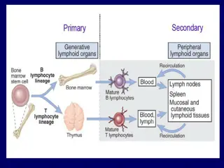
Diagram showing development of all the blood cells and how lymphocytes have a separate origin from other white blood cells. 39
Lymphocytes B and T Cells Remember, lymphocytes are one of the categories of white blood cells Each B or T cell has ~100,000 antigen receptors all of the exact same type Each B or T cell recognizes a single antigen The receptor molecules and recognition process are different for B cells vs. T cells Both types of receptors are protein-based Both have both constant and variable regions 40
Constant regions have stable amino acid sequences from cell to cell; Variable regions have different amino acid sequences from cell to cell Diagram showing the receptor molecules in B cells and T cells. This diagram is used several times in the next sequence of slides. 41
Antigen Recognition B Cells B cell receptors are Y-shaped Each branch of the Y has 2 parts, called chains Inner, heavy chain makes the full Y Outer, light chain is located on the branches of the Y Both chains are proteins Chains are linked by chemical bonds The bottom of the Y is anchored in the B cell membrane 42
The protein structure of a B cell receptor 44
Antigen Recognition B Cells The bottom regions of both chains have constant amino acid sequences The outer branches of both chains have variable amino acid sequences These variable ends are the antigen binding sites They bind directly to the epitopes B cells recognize unaltered antigens! 45
Antigen Recognition T Cells T cell receptors are unbranched chain and chain are chemically linked Both are anchored in the membrane Both have basal constant regions and terminal variable regions A single antigen binding site is at the terminus 47
T Cells DO NOT recognize intact antigens on intact pathogens T cells recognize antigen fragments that have been bound to a self-cell protein called a major histocompatibility molecule MHC major histocompatibility complex of genes codes for these molecules MHC molecules bind to antigen fragments inside a self-cell, and present the fragments at the surface of the cell T cells detect the presented antigen+MHC complex 49
MHC self-cell proteins Diagram showing the production of MHC molecules, how they become attached to antigen fragments, and how the complex is presented at the cell surface. This diagram is used repeatedly in the next sequence of slides. 50


 attacking a")