ANTENATAL FETAL ASSESSMENT
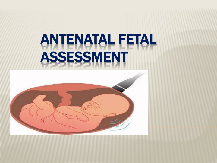
Antenatal fetal assessment is crucial for identifying and preventing potential risks to the fetus, such as neurologic injury or mortality. By evaluating fetal well-being, growth, movement, amniotic fluid levels, and lung maturity, healthcare providers can mitigate complications like stillbirth, metabolic acidosis, and cerebral palsy. Conditions associated with increased perinatal morbidity/mortality, such as small for gestational age fetuses and pre-eclampsia, highlight the importance of timely assessments. Starting fetal assessment antenatally, based on individual risk factors, is essential for optimal pregnancy management and maternal-fetal health.

Download Presentation

Please find below an Image/Link to download the presentation.
The content on the website is provided AS IS for your information and personal use only. It may not be sold, licensed, or shared on other websites without obtaining consent from the author.If you encounter any issues during the download, it is possible that the publisher has removed the file from their server.
You are allowed to download the files provided on this website for personal or commercial use, subject to the condition that they are used lawfully. All files are the property of their respective owners.
The content on the website is provided AS IS for your information and personal use only. It may not be sold, licensed, or shared on other websites without obtaining consent from the author.
E N D
Presentation Transcript
ANTENATAL FETAL ANTENATAL FETAL ASSESSMENT ASSESSMENT
OBJECTIVES: Describe how to test for each of the following Fetal well-being Fetal growth Fetal movement Amniotic fluid Fetal lung maturity - - - - -
FETAL ASSESSMENT ( FETAL ASSESSMENT (FETAL WELL FETAL WELL- -BEING) BEING) Fetal assessment is to identify fetuses at risk of neurologic injury or death in order to prevent it To prevent prenatal mortality & morbidity FETAL AND NEONATAL COMPLICATIONS OF FETAL AND NEONATAL COMPLICATIONS OF ANTEPARTUM ASPHYXIA ANTEPARTUM ASPHYXIA Stillbirth (Mortality) Stillbirth (Mortality) Metabolic acidosis at birth Metabolic acidosis at birth Hypoxic renal damage Hypoxic renal damage Necrotizing enterocolitis Necrotizing enterocolitis Intracranial hemorrhage Intracranial hemorrhage Seizures Seizures Cerebral palsy Cerebral palsy
RATIONAL If fetal oxygenation challenged: - blood flow directed to brain, heart & adrenal & blood flow away from the kidney fetal urine production decrease AF volume. - CNS hypoxia Fetal movement decrease -chemoreceptor's vegally-mediated reflex Fetal heart rate abnormality late deceleration. decrease
CONDITIONS ASSOCIATED WITH INCREASED CONDITIONS ASSOCIATED WITH INCREASED PERINATAL MORBIDITY/MORTALITY PERINATAL MORBIDITY/MORTALITY Small for gestational age fetus Decreased fetal movement Postdates pregnancy (>294 days) Pre-eclampsia/chronic hypertension Pre-pregnancy diabetes Insulin requiring gestational diabetes Preterm premature rupture of membranes Chronic (stable) abruption
WHEN TO START FETAL ASSESSMENT WHEN TO START FETAL ASSESSMENT ANTENATALLY ANTENATALLY ** Risk assessed individually **For D.M. fetal assessment should start from 32 weeks onward if uncomplicated ***If complicated D.M. start at 24 weeks onward **For Post date pregnancy start at 40 weeks **For any patient with decrease fetal movement start immediately ** Fetal assessment is done once or twice weekly
EARLY PREGNANCY ASSESSMENT Fetal heart activity fetal auscultation (special stethoscope or Doppler) ~12weeks Nuchal translucency measurement for early screening for chromosomal abnormality Between 11-13+ weeks
fetal heart activity seen by USS Can be seen from 6weeks
EARLY PREGNANCY ASSESSMENT Fetal movement Fetal movement are usually first perceptible to mother ~17w-20w (quickening) 50% of isolated limb movements are perceived 80% of trunk and limb movements Fetal growth Fundal height USS
FETAL GROWTH By fundal height measurement in the clinic By ultrasound Biometry Biometry: Biparietal diameter (BPD) Abdominal Circumference (AC) Femur Length (FL) Head Circumference (HC) Amniotic fluid Amniotic fluid
GROWTH CHART GROWTH CHART
ASSESSMENT OF FETAL GROWTH BY ULTRASOUND AC AC BPD BPD FL FL
LATE PREGNANCY ASSESSMENT Fetal movement counting kick chart Contraction stress test CST Non stress test NST Doppler Velocimetry UAV amniotic fluid index AFI
FETAL MOVEMENT COUNTING FETAL MOVEMENT COUNTING It should be started ~ It should be started ~28 &~ &~24 24w in high risk pregnancy w in high risk pregnancy It can reduce avoidable stillbirth It can reduce avoidable stillbirth CARDIFF TECHNIQUE CARDIFF TECHNIQUE - -10 10 movement in movement in 12 - -If abnormal patient should get further assessment If abnormal patient should get further assessment SADOVSKY TECHNIQUE SADOVSKY TECHNIQUE - -4 4 movement /hour if not felt another hour movement /hour if not felt another hour If not patient need more assessment If not patient need more assessment 28w in normal pregnancy w in normal pregnancy 12 hours hours
CONTRACTION STRESS TEST (CST) Causing uterine contraction over 20minutes At least 2 uterine contractions Uterine contraction restrict O2 delivery to the fetus Normal fetus will tolerate contraction Hypoxic fetus will have late deceleration High false positive rate ~50% 100% true negative rate
NON STRESS TEST (NST) Main advantage over CST is no need for contraction False +ve & false ve higher than CST done
NON STRESS TEST NON STRESS TEST The base line 120-160 beats/minute Different criteria in fetuses <32w Reactive Reactive: At least two accelerations from base line of 15 bpm for at least 15 sec within 20 minutes Non reactive Non reactive: No acceleration after 20 minutes- proceed for another 20 minutes
NON STRESS TEST ( NON STRESS TEST (NST) NST) If non reactive in 40 minutes---proceed for contraction stress test or biophysical profile The positive predictive value of NST to predict fetal acidosis at birth is 55%
INTERPRETATION OF CTG INTERPRETATION OF CTG Normal Baseline FHR 110 160 bpm Moderate bradycardia 100 109 bpm Moderate tachycardia 161 180 bpm Abnormal bradycardia < 100 bpm Abnormal tachycardia > 180 bpm
DECELERATION DECELERATION EARLY : Head compression LATE : U-P Insufficiency VARIABLE : Cord compression Primary CNS dysfunction
TACHYCARDIA TACHYCARDIA HYPOXIA HYPOXIA CHORIOAMNIONITIS MATERNAL FEVER B-MIMETIC DRUGS FETAL ANAEMIA,SEPSIS,HT FAILURE,ARRHYTHMIAS
AMNIOTIC FLUID VOLUME ~AFI Amniotic fluid index AFI -the sum of the maximum vertical fluid pocket diameter in four quarters -the normal value 5-25cm -<5~ oligohydraminous ->24cm polyhydraminous
BIOPHYSICAL PROFILE (BPP) Combines NST with USS estimation AFV, fetal breathing, body movement & reflex/tone/extension-flexion movement . it is a scoring system it is done over 30minute It measure acute hypoxia(NST, body mov. &breathing) & chronic hypoxia (AFI)
FETAL BIOPHYSICAL PROFILE/NST+ Normal (score=2) Biophysical Variable 1 episode FBM of at least 30 s duration in 30 min movements Abnormal (score= 0) Absent FBM or no episode >30 s in 30 min 2 or fewer body/limb movements in 30 min Fetal breathing Fetal movements 3 discrete body/limb movements in 30 min Fetal tone 1 episode of active extension with return to flexion of fetal limb(s) or trunk. Opening and closing of the hand considered normal tone Either slow extension with return to partial flexion or movement of limb in full extension Absent fetal movement Either no AF pockets or a pocket<2 cm in 2 perpendicular planes Amniotic fluid volume 1 pocket of AF that measures at least 2 cm in 2 perpendicular planes
BPP The risk of fetal death within 1 week if BPP is normal~ 1/1300 Modified BPP (mBPP) -NST & AFI -low false negative 0.8/1000 -high false positives ~60%
DOPPLER VELOCIMETRY Measurement of blood flow velocities in maternal & fetal vessels Reflect feto-placental circulation Doppler indices from UA, Uterine A & MCA Doppler studies is mostly valuable IUGR In IUGR absent or reversed EDF (end diastolic flow) associated with fetal hypoxia
INVASIVE FETAL ASSESSMENT Amniocentesis
AMNIOCENTESIS Obtaining a sample of amniotic flui during pregnancy. Obtaining a sample of amniotic flui during pregnancy. Usullay done after Usullay done after 15 15w (can be done after w (can be done after 11 Indication Indication -genitic (karyotype) genitic (karyotype) - -billirubin level (RH billirubin level (RH- -isimunisation) isimunisation) - -fetal lung maturity (L/S) fetal lung maturity (L/S) - -therputic in polyhydramnios therputic in polyhydramnios Risks: ROM ~ Risks: ROM ~1 1%, abortion %, abortion 0.5 11w) w) 0.5%, infection %, infection 1 1/ /1000 1000
CVS CHORIONIC VILLUS SAMPLING Usually done after 10w It is the procedure of choice for first trimester prenatal diagnosis of genetic disorders Complication: fetal loss (0.7 CVS procedure and CVS procedure and 1.3 1.3 percent within percent within 30 induced limb defects Second trimester amniocentesis is associated with the lowest risk of pregnancy loss; chorionic villus samplings safer than early (i.e, before 15 weeks) amniocentesis. 0.7 percent within percent within 14 30 days) 14 days of a TA days of a TA days), , Procedure- .
CORDOCENTESIS Indication: - rapid karyotyping -diagnosis of inherited disorders -fetal HB assessment -fetal plt level -fetal blood transfusion Complication: bleeding, bradycardia, infection .
FETAL DNA IN MATERNAL BLOOD -Used if indication by ultrasound for testing or history of genetic disease Result available in 2 weeks-
FETAL LUNG MATURITY A test for fetal lung maturity is performed before semi-elective but medically indicated births <39 weeks Tests for fetal lung maturity are generally not performed before 32 weeks of gestation RDS develops as a consequence of surfactant deficiency and immature lung development. L/S ratio is the most commonly used (ratio should be 2:1
-FLM TESTING MAY HAVE VALUE IN THE FOLLOWING CLINICAL SITUATIONS: -Premature rupture of membranes ( 32 weeks) if FLM test is mature, delivery is likely safer than wait and see approach Assessment of need for NICU possible only if early delivery has medical mandate and time allows for FLM testing Other selected late preterm and early preterm pregnancy issues where FLM may guide management of at-risk pregnancy
FETAL LUNG MATURITY FLM All tests require amniocentesis for obtaining amniotic fluid Comparison of FLM Laboratory Testing Options Lecithin-sphingomyelin ratio (L/S) Lamellar body count (LBC) Phosphatidylglycerol (PG) Main role is in adjudication of immature LBC or PG Last test of choice Labor intensive, imprecise Limited availability Results take >24 hrs unless performed at a local laboratory Initial FLM of choice Rapid, sensitive New data indicates that one can estimate risk of respiratory distress syndrome (RDS) as a function of gestational age and LBC Not useful unless gestational age 35 weeks Limited availability Sensitive


")
")


")