Antifungal Susceptibility Testing in Candida Species

The study focuses on identifying antifungal-resistant isolates in Candida species, particularly C. albicans, C. glabrata, C. parapsilosis, and C. tropicalis, obtained from various samples. The research aims to assess potential anatomical resistance compartmentalization and understand resistance rates to fluconazole and echinocandins.

Download Presentation

Please find below an Image/Link to download the presentation.
The content on the website is provided AS IS for your information and personal use only. It may not be sold, licensed, or shared on other websites without obtaining consent from the author.If you encounter any issues during the download, it is possible that the publisher has removed the file from their server.
You are allowed to download the files provided on this website for personal or commercial use, subject to the condition that they are used lawfully. All files are the property of their respective owners.
The content on the website is provided AS IS for your information and personal use only. It may not be sold, licensed, or shared on other websites without obtaining consent from the author.
E N D
Presentation Transcript
Antifungal Susceptibility Testing Identifies the Abdominal Antifungal Susceptibility Testing Identifies the Abdominal Cavity as a Source of Candida glabrata Cavity as a Source of Candida glabrata- -Resistant Isolates Resistant Isolates : : 2
INTRODUCTION INTRODUCTION 4
INTRODUCTION INTRODUCTION The likelihood of developing invasive candidiasis increases in at-risk patients with mucosal colonization by Candida species, a condition mostly caused by C. albicans,Candida parapsilosis, C. glabrata, or Candida tropicalis (1). Blood Culture Miscellaneous Clinical Samples Fluconazole Resistance Rates 0.3% (C. albicans) to 8% (C. glabrata) 9.2% (including 42% of vaginal isolates ) Echinocandin Resistance Rates 0.1% (C. albicans) to 3.5% (C. glabrata) 1.9% (including 7% of urine isolates ) Echinocandin resistance in nonblood Candida isolates suggests the existence of other anatomical reservoirs for resistant isolates (14, 15). 5
INTRODUCTION INTRODUCTION Here, we aim to: identify nonbloodstream antifungal-resistant isolates. potential anatomical resistance compartmentalization. We assessed antifungal susceptibility of a large number of isolates belonging to the four most common Candida spp. recovered from blood cultures, nonblood sterile samples, and nonsterile samples. 6
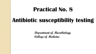
MATERIALS AND MATERIALS AND METHODS METHODS 7
MATERIALS AND METHODS MATERIALS AND METHODS Study period and isolates From July 1, 2016, to June 30, 2019 at the Clinical Microbiology and Infectious Diseases Department of Gregorio Mara n Hospital (Madrid, Spain) Selected isolates 1,103 7,767 Total isolates C. albicans 5,402 695 C. glabrata 1,150 182 C. parapsilosis 711 142 C. tropicalis 504 84 8
MATERIALS AND METHODS MATERIALS AND METHODS 9
MATERIALS AND METHODS MATERIALS AND METHODS Isolate identification, antifungal susceptibility testing, and genotyping Isolates were identified as routinely done in our laboratory (MALDI Biotyper); those with an antifungal non-wild-type phenotype, were further molecularly confirmed (29). Antifungal susceptibility to amphotericin B, fluconazole, voriconazole, and posaconazole, isavuconazole, micafungin, and anidulafungin was assessed by EUCAST E.Def 7.3.2 broth dilution method using tissue-treated plates (30). Isolates were categorized as susceptible or resistant according to EUCAST clinical breakpoints and as wild type or non- wild type based on epidemiological cutoff values (ECOFFs) or wild type upper limits for isavuconazole (31, 32). Echinocandin or fluconazole non-wild-type isolates were retested, submitted for fks or erg11 gene sequencing (5, 33), and further genotyped by using species-specific microsatellite markers to detect C. albicans, C.parapsilosis, C. glabrata and C. Tropicalis in order to detect non-wild-type clones spreading across the hospital. 10
MATERIALS AND METHODS MATERIALS AND METHODS Isolate identification, antifungal susceptibility testing, and genotyping Isolates were considered to have identical genotypes when they showed the same alleles in all loci (28, 34). We defined resistance compartmentalization in a given patient as the presence of identical genotypes showing a resistant phenotype in one compartment and a susceptible phenotype in another(s). 11
RESULTS RESULTS 12
RESULTS RESULTS Isolate species and clinical sources groups patients group 1 (blood) 54 group 2 (other normally sterile sites) 139 groups 1 and 2 11 group 3 (nonsterile sites) 133 group 4 (nonsterile sites) 170 Overall 507 13
RESULTS RESULTS Isolate species and clinical sources 14
RESULTS RESULTS Antifungal susceptibility and molecular characterization of non-wild-type isolates No resistance to amphotericin B 16 of 1,103 isolates (1.5%) were fluconazole-resistant, mostly C. glabrata. (C. glabrata , n = 13 ; C. albicans , n = 2 ; C. Tropicalis , n = 1) 2.5% (n = 28) of the isolates were fluconazole non-wild type: C. glabrata (n = 13), C. albicans (n = 10), and C. Tropicalis (n = 5) Panazole resistance was detected in 11 isolates. (C. glabrata, n = 10; C. albicans, n = 1) The percentages of non-wild-type isolates tovoriconazole, posaconazole, and isavuconazole were 1.3% (n = 14), 1.0% (n = 11), and 1.4% (n = 15), respectively .all were fluconazole non-wild type as well. 14 of 1,103 (1.3%) isolates were echinocandin-resistant, mostly C. glabrata (n = 10). Eleven isolates (C. glabrata [n = 8], C. tropicalis [n = 2], and C. albicans [n = 1]) were micafungin and anidulafungin non-wild-type, three C. albicans were micafungin non-wild-type, two C. glabrata isolates were anidulafungin non-wild-type, and one C.parapsilosis isolate was micafungin non-wild- type. 1.5% (n = 17) of the isolates were echinocandin non-wild- type. C. Glabrata (n = 10), C. albicans (n = 4), C. tropicalis (n = 2), and C. parapsilosis (n = 1). 15
RESULTS RESULTS Description of patients with resistant isolates 16
RESULTS RESULTS Description of patients with resistant isolates Description of patients with resistant isolates gender 67% male (33 % female) median age 61 years mortality 67% Clinical presentation candidemia (n = 3), candidemia and intra-abdominal candidiasis (n = 4), candidemia and endocarditis (n = 1), intra-abdominal candidiasis (n = 5), and other forms of candidiasis (n = 2) most common risk factors antibiotic use (100%), intravascular catheter (73%), urinary catheter (60%), parenteral nutrition (53%), and abdominal surgery (60%). Previous Antifungal use From the patients carrying fluconazole-resistant isolates, five (63%) had previously used azoles; likewise, of the nine patients carrying echinocandin-resistant isolates, eight (89%) had been previously treated with echinocandins. Nine patients were on antifungal treatment with echinocandins (n = 6) or fluconazole (n = 3) at the moment of resistant isolate collection, with 78% mortality. 17
RESULTS RESULTS Antifungal resistance in different anatomical compartments and compartmentalization of resistance. nonsterile samples (n = 9; 1.4%) blood cultures (n = 1; 0.7%) fluconazole resistance nonblood sterile samples (n = 6; 2.1%) echinocandin resistance nonblood sterile samples (n = 7; 2.4%) nonsterile samples (n = 5; 0.8%) blood cultures (n = 2; 1.3%) 18
RESULTS RESULTS Antifungal resistance in different anatomical compartments and compartmentalization of resistance. Fluconazole/echinocandin resistance rates studied compartments bloodstream (0.7%/1.3%) abdominal cavity (3.2%/3.2%) deep organs (0.9%/1.9%) We found statistically nonsignificant higher percentages of fluconazole-resistant isolates (3.2% versus 0.7%) and echinocandin-resistant isolates (3.2%versus 1.3%) in abdominal cavity samples than in isolates from blood cultures. deep-seated soft tissues (0%/0%) skin and mucosa (1%/0.5%) lower respiratory tract (1%/0.7%) urinary tract (3.1%/1.6%) other nonsterile sources (0%/0%) 19
RESULTS RESULTS Antifungal resistance in different anatomical compartments and compartmentalization of resistance. C. Glabrata isolate of abdominal cavity samples C. Glabrata isolate of blood cultures fluconazole-resistant C. Glabrata isolates 11.9% 3.2% echinocandin-resistant C. Glabrata isolates 7.1% 3.2% C. Glabrata was the only species for which resistant isolates were found in all isolate groups the rate of fluconazole resistance (11.9% / 3.2%, P . 0.05) and echinocandin resistance (7.1% / 3.2%) for C. Glabrata, tended to be higher in isolates from the abdominal cavity than in isolates from blood cultures, respectively. 20
RESULTS RESULTS We found compartmentalization of antifungal resistance in 6 of 15 patients (40%) with different forms of invasive candidiasis; isolates were mostly C. glabrata and led to a mortality of 67% (4 of 6). (Patient timelines are shown in Fig. 2.) One patient harbored a fluconazole- resistant isolate, four patients harbored echinocandin-resistant isolates,and one patient harbored a fluconazole- and echinocandin-resistant isolate. Isolates were sourced from two (patients 8, 10, and 14), three (patient 15), or four (patients 13 and 16) compartments (Table 3). 21
RESULTS RESULTS Antifungal resistance in different anatomical compartments and compartmentalization of resistance. Resistant isolates were collected during (n = 5) or after (n = 6) antifungal treatment and were mostly echinocandin- resistant C. glabrata isolates from the abdominal cavity. Additionally, we found antifungal resistance compartmentalization in patients 1 and 9 with nonsignificant isolates (Table 3). In the remaining nine patients infected by resistant isolates, compartmentalization of resistance could not be ruled out because: only one isolate per patient was studied (n = 5), isolates from different compartments proved to have different genotypes (n = 1), Coexistence of resistant isolates and susceptible isolates with the same genotype occurred in samples from the same compartment (n = 2), and isolates with identical genotypes from the same compartment showed resistance (n = 1). 22
DISCUSSION DISCUSSION 23
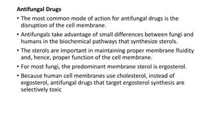
DISCUSSION DISCUSSION Non-wild type is a concept based on epidemiological cutoff values (ECOFFs),whereas resistance is based on clinical breakpoints. ECOFFs and clinical breakpoints are identical in most of cases, with the exception of azoles against C. albicans and C.tropicalis. We found no erg11 gene mutations concerning exclusively fluconazole-resistant C. albicans isolates (17, 18). In contrast, the presence of erg11 mutations may play a role in the promotion of fluconazole resistance in C. tropicalis as shown by the existence of a previously described mutation (G464S) (19) in a fluconazole-resistant C. Tropicalis isolate and the same mutation in heterozygosis (G464S/G) in a non-wild-type isolate. Antifungal resistance was mostly dominated by C. glabrata (n = 20/27 resistant isolates). Most resistant isolates found in this study were C. glabrata from the abdominal cavity. Echinocandin-resistant isolates harbored previously reported fks gene mutations, except the R1361H substitution in a C. albicans isolate from patient 8 (5, 6, 20,21). An alternative substitution at the same position (R1361L) has been previously linked to a highly caspofungin-resistant phenotype (22). Resistance was not caused by the spread of resistant clones but by prior antifungal use. Overall, resistance rates tended to be lower in isolates from blood cultures than in isolates from other compartments, which is in line with our previous observations (5). Resistance compartmentalization illustrates how resistance might be overlooked if susceptibility testing is restricted to bloodstream isolates. 24
DISCUSSION DISCUSSION In vitro secondary acquisition of echinocandin resistance in C. glabrata has been associated with prior echinocandins exposure (23). In fact, we identified 15 patients with different forms of candidiasis, and in 12 of them the resistant isolates (8 with echinocandin-resistant isolates) were collected after or during antifungal treatment. The presence of echinocandin-resistant C. glabrata isolates in the abdominal cavity may be caused by subinhibitory echinocandin concentrations due to restrained diffusion to the peritoneum (24, 25), not observed with fluconazole (26). Alternatively, C. Glabrata isolates in the gut may become echinocandin-resistant before translocation during treatment, given that echinocandins are excreted unaltered through the gut (27). The abdominal cavity can be considered a reservoir for resistant isolates (14 16). Antifungal resistance compartmentalization was found in six patients, mostly harboring echinocandin-resistant C. glabrata. With the exception of one patient, the remaining five had susceptible isolates in blood cultures, whereas resistant isolates originated from other compartments, mostly the abdominal cavity. Although it is difficult to discern the role of resistance in the outcome, high mortality was seen (67%). Three genotypes clustered in two (CG-1) and three patients (G-6 and CG-24).Given that they were epidemiologically unrelated, promotion of resistance seems to have occurred intrapatient after antifungal treatment and not because of the spread of resistant clones. Genotypes CG-6 and CG-24 are frequently detected and found in either antifungal-resistant or antifungal-susceptible isolates, suggesting a certain ability of both genotypes to acquire antifungal resistance(28). 25
DISCUSSION DISCUSSION Our study is subjected to limitations. The retrospective nature prevented the collection of additional isolates in patients harboring resistant isolates; moreover, antifungal resistance compartmentalization would have been detected in more patients if a more systematic prospective collection had been done. Finally, assessing the role of resistance in the prognosis of patients would have involved comparisons with noncompartmentalized patients, which was out of the scope of the study. 26
Thanks For Your Attention 27