Approach to Heart Murmur: Understanding Signs & Identification
This informative guide explains the basics of heart murmurs, the significance of identifying pathological murmurs, clues for identification, and the physiology behind heart murmur sounds. Learn how to approach patients with heart murmurs effectively.

Download Presentation

Please find below an Image/Link to download the presentation.
The content on the website is provided AS IS for your information and personal use only. It may not be sold, licensed, or shared on other websites without obtaining consent from the author.If you encounter any issues during the download, it is possible that the publisher has removed the file from their server.
You are allowed to download the files provided on this website for personal or commercial use, subject to the condition that they are used lawfully. All files are the property of their respective owners.
The content on the website is provided AS IS for your information and personal use only. It may not be sold, licensed, or shared on other websites without obtaining consent from the author.
E N D
Presentation Transcript
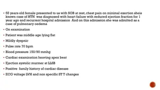
APPROACH TO A PATIENT WITH HEART MURMUR DR AMRITA KURIAN JUNIOR RESIDENT DEPT OF GENERAL MEDICINE
BASICS S1 : CLOSURE OF AV VALVES S2 : CLOSURE OF SEMILUNAR VALVES SYSTOLE : PERIOD BETWEEN S1 AND S2 BLOOD FLOWS ACROSS AORTIC AND PULMONARY VALVES DIASTOLE : PERIOD BETWEEN S2 AND S1 BLOOD FLOWS ACROSS MITRAL AND TRICUSPID VALVES
SIGNIFICANCE OF A MURMUR When someone is found to have a murmur, the first question to ask is whether the murmur is organic (pathologic), or does it represent a normal variant. A "yes" answer indicates the presence of structural cardiovascular disease. Proceed to cardiac physical examination, to determine the likeliest hemodynamic cause of the murmur and the anatomic origin. Next, the possible etiologies of the heart disease should be investigated, and thus an assessment of the severity of the underlying condition can be made.
CLUES TO IDENTIFY PATHOLOGICAL MURMURS All diastolic murmurs. All pansystolic and late systolic murmurs. Continuous murmurs. Very loud murmurs (grade 4 or higher) Associated cardiac abnormalities (presence of left ventricular hypertrophy on precordial palpation, an ejection or midsystolic click, or an opening snap would suggest that the heart murmur represents a cardiac structural abnormality)
PHYSIOLOGY Heart murmurs are caused by audible vibrations that are due to increased turbulence from accelerated blood flow through normal or abnormal orifices. flow through a narrowed or irregular orifice into a dilated vessel or chamber, or If blood flows in an abnormal direction; say (1)backward flow through an incompetent valve (2) ventricular septal defect, or (3) patent ductus arteriosus.
Thus, turbulence is thought to be the primary factor in the genesis of heart murmurs. Determinants of turbulence : the velocity of blood flow; increase in velocity result in marked increase in turbulence. An obstruction to blood flow, like stenosis produces abnormal turbulence. Turbulence increases as the pressure gradient across the valve becomes larger. Turbulence increases when blood moves from a small to large chamber. Low blood viscosity (e.g., anemia) enhances turbulence.
CHARACTERISITCS OF A HEART MURMUR The following features of murmurs should be consciously analyzed in each patient: Whether the murmur is systolic or diastolic The timing of the murmur within systole or diastole : early, mid, or late The duration of the murmur The intensity or loudness of the murmur, e.g., its grade The location and radiation of murmur The shape and pitch of the murmur Other cardiac abnormalities present on examination that may relate to the etiology of the murmur.
TIMING Identify S1 and S2. Palpating the carotid pulse helps to determine the onset of ventricular systole. The carotid upstroke closely follows S1. Systolic murmurs begin with or after the first heart sound (S1) and terminate at or before the component (A2 or P2) of the second heart sound (S2) that corresponds to their site of origin. Diastolic murmurs begin with or after the associated component of S2 and end at or before the subsequent S1. Continuous murmurs are not confined to either phase of the cardiac cycle but instead begin in early systole and proceed through S2 into all or part of diastole.
Within systole: Early systolic Mid systolic Late systolic Holocystolic Within diastole Early diastolic Mid- diastolic
DURATION SYSTOLE The duration of a heart murmur depends on the length of time over which a pressure difference exists between two cardiac chambers, the left ventricle and the aorta, the right ventricle and the pulmonary artery, or the great vessels. Any murmur that is truly holo- or pansystolic, regardless of shape, is caused by continuous pressure differential between two cardiac chambers. Thus, the classic murmurs of a ventricular septal defect or mitral regurgitation begin with S1 and extend through S2. Severe obstruction of the left or right ventricular outflow tract (severe Aortic or Pulmonary stenosis) may cause a prolonged systolic murmur that is actually pansystolic.
The presence of a long systolic murmur should stimulate a careful evaluation of late systole through the use of selective listening. A careful focus on the last 25% of systole is important. If sound vibrations end before S2 ie spares the last 1/3rdof systole, the murmur is usually an ejection murmur. If the murmur extends to S2 (pansystolic), the differential diagnosis should include regurgitant lesions as well as severe semilunar valve stenosis.
DIASTOLE Flow across a nonobstructive or mildly stenotic A-V valve produces a short mid- diastolic murmur A long diastolic murmur following an opening snap is indicative of a persistent A-V gradient and significant mitral stenosis. With short cycle lengths, these murmurs will extend to S1. Murmurs of aortic and pulmonary regurgitation start in early diastole and extend into mid-diastole.
INTENSITY grade 1 very soft heard with great effort grade 2 easily heard grade 3 loud but without thrill grade 4 loud with thrill grade 5 heard with stethoscope edge touching the chest (thrill present) grade 6 heard with stethoscope slightly off the chest Grade 3 or more indicates a structural heart disease .
It also implies high blood flow velocity at the site of murmur production. (eg: Small VSDs are accompanied by loud; usually grade 4 or greater systolic murmurs ) Factors diminishing intensity: obesity. obstructive lung disease. a large pericardial effusion. when cardiac output is reduced significantly. when the pressure gradient between the involved cardiac structures is low.
Increased Intensity High cardiac output (hyperdynamic) states Thin chest wall Narrow thoracic diameter, e.g., "straight back," pectus excavatum Anemia (decreased blood viscosity) Tortuous aorta (close to chest wall)
LOCATION AND RADIATION Recognition of the location and radiation of the murmur help facilitate its accurate identification. Adventitious sounds, such as a systolic click or diastolic snap, or abnormalities of S1 or S2 may provide additional clues. SHAPE Crescendo Crescendo-decrescendo : eg; Aortic stenosis Decrescendo: eg chronic Aortic regurgitation
Plateau: chronic Mitral regurgitation PITCH HIGH WHISTLE LOW- RUMBLE High pitched murmurs often correspond to high pressure gradients, hence the diastolic murmur of Aortic regurgitation (AR) is high pitched whereas murmur of mitral stenosis (MS) is a low frequency event. The coarse systolic murmur of aortic stenosis (AS) may sound higher pitched and more acoustically pure at the apex :Gallavardin effect.
EARLY SYSTOLIC MURMURS Begin with S1,extend for a variable period and end well before S2. 1) Acute severe MR Regurgitation into a relatively non compliant normal sized atrium leads to an early, decrescendo murmur best heard at or just medial to apex. Causes are papillary muscle rupture after acute MI, rupture of chordae tendinae in MVP, Infective endocarditis and Blunt chest wall trauma. 2) Small muscular VSD defect closes progressively during septal contraction. localised to left sternal border grade 4 or 5
3) Acute severe TR with normal pulmonary artery pressure Best heard at lower left sternal border soft: grade 1 or 2 increase in intensity with respiration (Carvallo s sign)
MID SYSTOLIC MURMURS Begin a short interval after S1 and end before S2 Crescendo-decrescendo in configuration Eg : 1 AORTIC STENOSIS Loudest at aortic area radiates to carotids. Gallavardin Effect transmission to apex where it becomes high pitched.
Features of severe AS: with a normal cardiac output; grade 4 or more murmur. soft or absent A2. paradoxical splitting of S2. apical S4 late peaking systolic murmur. Pulse: parvus et tardus
2) HOCM lower left sternal border The intensity of the murmur may vary from beat to beat and after provocative maneuvers but usually does not exceed grade 3. The murmur classically will increase in intensity with maneuvers that result in increasing degrees of outflow tract obstruction, such as a reduction in preload or afterload (Valsalva, standing, vasodilators), or with an augmentation of contractility (inotropic stimulation). Maneuvers that increase preload (squatting, passive leg raising, volume administration) or afterload (squatting, vasopressors) or that reduce contractility ( -adrenoreceptor blockers) decrease the intensity of the murmur.
3) PULMONARY STENOSIS pulmonic area (2ndand 3rdleft intercostal space) 4)Significant left to right shunting due to ASD Increased pulmonary blood flow and grade 2-3 midsystolic murmur at upper left sternal border with fixed split of S2 5)AORTIC VALVE SCLEROSIS 2ndright intercostal space, carotid upstrokes being normal and no LVH in ECG.
6) PHYSIOLOGIC MURMURS/ FUNCTIONAL MURMURS Flow or Functional Murmur: These are by far the most common heart murmurs and are found in many normal people. In the presence of a normal resting cardiac output the murmur is known as an innocent or functional murmur.
If the murmur is related to a hyperkinetic state with increased cardiac output and stroke volume, the murmur is known as a physiologic murmur. Eg: in pregnancy, hyperthyroidism or anaemia . Grade 1-2 mid systolic murmur heard the left lower sternal border. STILL S MURMUR: Benign, grade 2, vibratory or musical midsystolic murmur at the mid or lower left sternal border in normal children and adolescents, best heard in the supine position.
LATE SYSTOLIC MURMURS Best heard at left ventricular apex - Mostly due to MVP murmur is introduced by a non ejection click. With the posterior leaflet prolapse MR jet is directed anteriorly and medially - murmur radiates to the base of heart and masquerades AS. Anterior leaflet prolapse - posteriorly directed MR jet - radiates to axilla or left infrascapular region.
HOLOSYSTOLIC MURMURS Begin with S1, continue through systole to S2 1) Chronic MR Best heard at left ventricular apex, high pitched, plateau in configuration and radiates to the axilla. Causes of chronic MR : Rheumatic scarring of leaflets, Mitral annular calcification,post myocardial infarction LV remodelling, severe LV enlargement (dilated cardiomyopathy) 2) Chronic TR Softer than that of MR, loudest at lower left sternal border, Carvallo s sign positive.
Associated with c-v waves in JVP, pulsatile and enlarged liver, ascites and peripheral edema TR is more commonly a passive process. Causes of primary TR : Endocarditis, RHD, Carcinoid, Ebsteins Anomaly. 3) VSD Loudest at mid to lower left sternal border Radiates widely, thrill present in majority No change with inspiration
EARLY DIASTOLIC MURMURS Begin immediately after S2 and end well before S1, Decrescendo in configuration 1) Chronic AR High pitched, blowing , second right intercostal space. Auscultated with patient leaning forward at end expiration. With primary valve disease, murmur radiated along left sternal border. When AR IS due to aortic root disease, murmur may radiate along right sternal border. causes Marfans syndrome with aneurysm, annuloaortic ectasia, ankylosing spondylitis, aortic dissection
Chronic, severe AR also may produce a lower-pitched, mid to late, grade 1 or 2 diastolic murmur at the apex (Austin Flint murmur) Distinguished from MS by absence of opening snap and response to vasodilator challenge 2) Pulmonic Regurgitation decrescendo, early to mid-diastolic murmur (Graham Steell murmur) that begins after the pulmonic component of S2 Best heard at the second left interspace, and radiates along the left sternal border.
The intensity of the murmur may increase with inspiration. PR is most commonly due to dilation of the valve annulus from chronic elevation of the pulmonary artery pressure. Signs of PAH (left parasternal heave and loud, single or narrow split S2 are present) PR can also occur in endocarditis, with congenitally deformed valve or after repair of TOF.
MID DIASTOLIC MURMURS Produced due to obstruction/augmented flow at mitral or tricuspid valve Eg : 1 Mitral stenosis Most common cause rheumatic fever opening snap shortly after S2, followed by a low pitched murmur best heard at LV apex with patient in left lateral decubitus position. Presystolic accentuation which disappears in atrial fibrillation. The interval between A2 and opening snap varies inversely with the severity of stenosis.
2) Tricuspid stenosis lower left sternal border, increase in intensity with inspiration. prolonged y descent in JVP 3) Atrial Myxoma may prolapse across mitral valve and obstruct LV inflow. No opening snap or presystolic accentuation 4) Carey coombs murmur short mid diastolic murmur in Acute Rheumatic Fever, due to flow through edematous mitral valve.
CONTINUOUS MURMURS Begin in systole, peak near the second heart sound and continue into all or part of diastole PDA best heard at upper left sternal border Ruptured sinus of Valsalva aneurysm upper right sternal border. Coronary arteriovenous fistula. At AV fistula for hemodialysis. Over intercostal collaterals in patients with coarctation of aorta venous hum in children and young adults (in right supraclavicular fossa). Mammary souffle in pregnancy.
TO AND FRO MURMUR in AS + AR (Here the midsystolic component decrescendos as it approaches S2)
DYNAMIC AUSCULTATION RESPIRATION Left-sided murmurs may be best heard at end expiration, when lung volumes are minimized and the heart and great vessels are brought closer to the chest wall. This phenomenon is characteristic of the murmur of AR. Murmurs of right-sided origin, such as tricuspid or pulmonic regurgitation, increase in intensity during inspiration.
ALTERATIONS OF SYSTEMIC VASCULAR RESISTANCE The systolic murmurs of AR, MR and VSD become louder during sustained handgrip, simultaneous inflation of blood pressure cuffs on both upper extremities to pressures 20 40 mmHg above systolic pressure for 20 s, or infusion of a vasopressor agent. (All these manuever raise the aortic pressure) Murmur associated with AS or HOCM become softer or remain unchanged with these maneuvers
CHANGES IN VENOUS RETURN The majority of murmurs decrease in intensity during the strain phase of the Valsalva maneuver or standing (positive intrathoracic pressure and decreased venous return) Two notable exceptions are the murmurs associated with MVP and obstructive HOCM, both of which become louder during the Valsalva maneuver. Squatting which increases venous return increases the intensity of all murmurs except MVP and HOCM.
STRATEGY FOR EVALUATING HEART MURMURS


 Acute severe TR with normal pulmonary artery")


 HOCM –")
 PULMONARY STENOSIS")
 PHYSIOLOGIC MURMURS/ FUNCTIONAL MURMURS")


 Tricuspid stenosis")