Approach to Pleural Effusion: Understanding Causes and Mechanisms

Pleural effusion is an abnormal collection of fluid in the pleural space, typically caused by underlying diseases such as heart failure, pneumonia, malignancy, or pulmonary embolism. Various mechanisms, including altered permeability and reduced drainage, contribute to its development. This condition requires a comprehensive approach for accurate diagnosis and management.

Download Presentation

Please find below an Image/Link to download the presentation.
The content on the website is provided AS IS for your information and personal use only. It may not be sold, licensed, or shared on other websites without obtaining consent from the author.If you encounter any issues during the download, it is possible that the publisher has removed the file from their server.
You are allowed to download the files provided on this website for personal or commercial use, subject to the condition that they are used lawfully. All files are the property of their respective owners.
The content on the website is provided AS IS for your information and personal use only. It may not be sold, licensed, or shared on other websites without obtaining consent from the author.
E N D
Presentation Transcript
Approach to Pleural Effusion Dr Abdalla Elfateh Ibrahim Consultant & Assisstant Professor of Pulmonary Medicine King Saud University
Pleural effusions Abnormal collection of fluid in the pleural space resulting from excess fluid production or decreased absorption
Pleural effusions A common medical problem is an indicator of an underlying acute or chronic disease - pulmonary or - nonpulmonary with more than 50 recognized causes
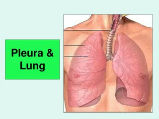
Anatomy The pleural space role in respiration 1- coupling the movement of the chest wall with that of the lungs in . A- a relative vacuum in the space keeps the visceral and parietal pleurae in close proximity. B- lubricant
Anatomy Pleural fluid volume 0.13 mL/kg/B. w (approximately 1 ml) Volume is maintained through the balance of - hydrostatic - oncotic pressure - lymphatic drainage
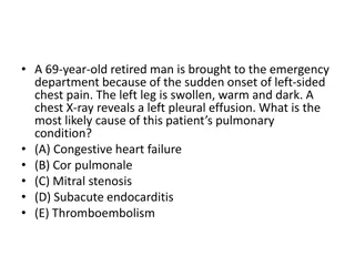
Etiology The etiologic spectrum of Pl.Ef is extensive most pleural effusions are caused by - congestive heart failure - pneumonia - malignancy - or pulmonary embolism.
Mechanisms Altered permeability of the pleural membranes Reduction in intravascular oncotic pressure Increased capillary hydrostatic pressure in the systemic and/or pulmonary circulation Decreased lymphatic drainage or complete blockage, including thoracic duct obstruction or rupture
Increased capillary permeability or vascular disruption (trauma, malignancy, inflammation, infection, pulmonary infarction, drug hypersensitivity, uremia, pancreatitis) Reduction in intravascular oncotic pressure (eg, hypoalbumiaemia ,cirrhosis) Increased capillary hydrostatic pressure in the systemic and/or pulmonary circulation ( congestive heart failure, superior vena cava syndrome) Decreased lymphatic drainage or complete blockage, including thoracic duct obstruction or rupture ( malignancy, trauma)
Types of pleural effusions Transudates pleural fluid proteins < 30 OR Exudates pleural fluid proteins >30
Causes of pleural effusion Transudates (usually bilateral effusion) Very Common causes Heart failure Liver cirrhosis
Transudates Less Common causes Hypoalbuminaemia Nephrotic syndrome Peritoneal dialysis Hypothyroidism Mitral Stenosis Myxedema Constrictive pericarditis Urinothorax (due to obstructive uropathy)
Causes of pleural exudates Common causes (Usually unilateral effusion) Parapneumonic effusions Malignancy ( lung or breast , lymphoma, leukemia; less commonly, ovarian carcinoma, stomach, sarcomas, melanoma, mesothelioma ) Tuberculosis Pulmonary embolism Collagen-vascular conditions (rheumatoid & systemic lupus erythematosus
Exudates Less Common causes Benign Asbestos-related effusion Pancreatitis Post-myocardial infarction Post CABG Pericardial disease Meigs syndrome (benign pelvic neoplasm with associated ascites and p.effusion) Ovarian hyperstimulation syndrome Drug-induced pleural disease Yellow nail syndrome (yellow nails, lymphedema, pleural effusions) Uremia Chylothorax (acute illness with elevated triglycerides in pleural fluid
Prognosis Varies in accordance with the condition s underlying etiology. The most common associated malignancy in men is lung cancer the most common associated malignancy in women is breast cancer malignant pleural effusion is associated with a very poor prognosis
Morbidity and mortality Related to - cause - stage of disease at the time of presentation - biochemical findings in the pleural fluid Parapneumonic effusion - Early diagnosis and treatment have a lower rate of complications -when treated promptly resolve without significant sequelae. - untreated or inappropriately treated parapneumonic effusions may lead to empyema, constrictive fibrosis, and sepsis
Clinical presentation Symptoms Asymptomatic Breathlessness Chest pain Cough Fever
Additional symptoms May suggest the underlying disease process. Example: lower limb oedema in heart failure Night sweats, fever, hemoptysis, and weight loss should suggest TB Hemoptysis also raises the possibility of malignancy
Clinical presentation History Infection, malignancy , risk of PE , heart failure etc.) Drug history An occupational history Asbestos exposure
Physical Examination Depend on the volume Dullness to percussion decreased tactile fremitus Asymmetrical chest expansion Mediastinal shift away from the effusion Signs of associated disease ( chronic liver disease-heart failure-nephrotic syndrome -SLE-RA-Ca lung)
DIAGNOSIS CXR Pleural aspiration CT scan Chest Ultrasound Pleural biopsy Medical thoracoscopy VAT (video assisted thoracoscopy) Bronchoscopy
Diagnostic Thoracentesis Pleural aspiration is a safe procedure in presence of sufficient fluid Contraindications - small volume of fluid - bleeding diathesis or anticoagulation - cutaneous disease over the puncture site Complication - pain at the puncture site - Bleeding - pneumothorax - Empyema - spleen/liver puncture.
Normal pleural fluid characteristics Straw colour (yellow) A pH of 7.60-7.64 Protein content of less than 2% (1-2 g/dL) Fewer than 1000 white blood cells (WBCs) per cubic millimeter Glucose content similar to that of plasma Lactate dehydrogenase (LDH) less than 50% of plasma
LABORATORY Biochemstry (5 ml) PH, proteins ,glucose and LDH send serum sample at the same time Microbiology lab (20ml) Gram stain + culture and sensitivity TB AFB &TB culture - PCR for TB Pathology lab (100ml or more) Looking for malignant cells Ask also for cell block(pleural clot)
Distinguish transudates from exudates Exudative effusions might be suspected by : ( observing the gross characteristics of the fluid ) Frankly purulent fluid indicates an empyema A milky fluid suggests a chylothorax (lymphatic obstruction) Grossly bloody fluid - trauma - malignancy, - postpericardiotomy syndrome - asbestos-related effusion (hematocrit of > 50% of the peripheral hematocrit defines a hemothorax)
Distinguish transudates from exudates Laboratory testing helps to distinguish pleural fluid transudates from Exudative Pleural fluid protein level greater than 2.9 g/dL
Distinguishing Transudates From Exudates The fluid is considered an exudate if any of the following applies:(Light`s criteria) Ratio of pleural fluid to serum protein greater than 0.5 Ratio of pleural fluid to serum LDH greater than 0.6 Pleural fluid LDH greater than two thirds of the upper limits of normal serum value
Distinguishing Transudates From Exudates Clinical judgment is required when pleural fluid test results fall near the cutoff points Example Heart failure on diuretics pleural fluid levels of N-terminal pro-brain natriuretic peptide (NT-proBNP) are elevated in effusions due to congestive heart
Distinguishing Transudates From Exudates serum - pleural protein = less than 3.1 g/dL s albumin pl. f. albumin = less than 1.2 g/dL
Pleural fluid pH Correlated with pleural fluid glucose levels Parapneumonic effusions a low fluid pH level is more predictive of complicated effusions fluid pH of less than 7.1-7.2 indicates the need for urgent drainage of the effusion
Pleural fluid glucose A low pleural glucose (30-50 mg/dL) suggests malignant effusion tuberculous pleuritis esophageal rupture lupus pleuritis. A very low pleural glucose (ie, < 30 mg/dL) suggest rheumatoid pleurisy or empyema.
Pleural Fluid Cell Count Differential Fluid lymphocytosis (> 85%) suggests -TB, lymphoma, sarcoidosis, rheumatoid , yellow nail syndrome, or chylothorax. Fluid lymphocyte ( 50-70% ) suggest malignancy
TB pleural effusion history of exposure positive PPD lymphocytic exudative effusions Pleural fluid adenosine deaminase Interferon-gamma test PCR for TB
IMAGING Ultrasonography Chest ultrasound is important for - Diagnosis of pleural effusion - guide aspiration and biopsy procedures CT Scanning Chest CT scanning with contrast
Biopsy should be considered, especially if TB or malignancy is suggested closed-needle p. biopsy is a blind technique ( bedside test) Medical thoracoscopy has a higher diagnostic yield Video assisted thoracoscopy
Treatment Treatment of the underlying medical disorder
Treatment Therapeutic Thoracentesis Remove larger amounts of pleural fluid is used to alleviate dyspnea
Treatment Parapneumonic effusions drainage (1) frankly purulent fluid, (2) a pleural fluid pH of less than 7.2 (3) loculated effusions (4) bacteria on Gram stain or culture Appropriate antibiotic treatment.
Treatment Tuberculous pleuritis typically is self-limited Disappear after treatment of TB Chylous effusions usually managed by dietary and surgical proc.
Treatment Malignant pleural effusions drain large, malignant PE to relieve dyspnea pleurodesis for recurrent effusions or placement of indwelling tunneled catheters.(eg, PleurX cathiter)
Treatment Pleurodesis (also known as pleural sclerosis) instilling an irritant into the pleural space to cause inflammatory changes that result in bridging fibrosis between the visceral and parietal pleural surfaces Sclerosing agents talc, doxycycline, bleomycin
Surgical treatment Video-assisted thoracoscopy to drain loculated pleural fluid obliterate the pleural space. Surgically implanted pleuroperitoneal shunts