Arthrodesis of DIJ and DIP Joints in Cattle
Learn about the intra-operative procedures for arthrodesis of the distal interphalangeal joint (DIJ) and distal interphalangeal joint (DIP) in cattle, including surgical techniques, post-operative care, and images illustrating the process. Understand the surgical approaches, anesthesia methods, and aftercare protocols involved in these joint fusion procedures.
Download Presentation

Please find below an Image/Link to download the presentation.
The content on the website is provided AS IS for your information and personal use only. It may not be sold, licensed, or shared on other websites without obtaining consent from the author. If you encounter any issues during the download, it is possible that the publisher has removed the file from their server.
You are allowed to download the files provided on this website for personal or commercial use, subject to the condition that they are used lawfully. All files are the property of their respective owners.
The content on the website is provided AS IS for your information and personal use only. It may not be sold, licensed, or shared on other websites without obtaining consent from the author.
E N D
Presentation Transcript
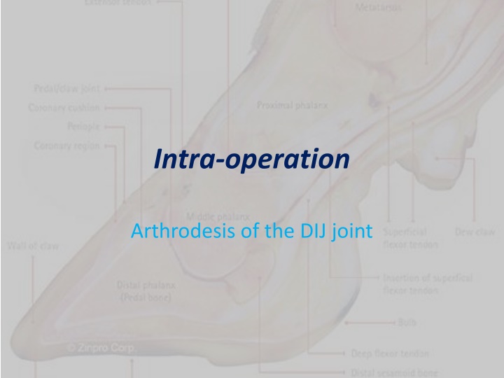
Intra-operation Arthrodesis of the DIJ joint
Arthrodesis of the DIP joint by the Solar Approach This procedure is performed under sedation and intravenous regional anaesthesia. The cattle is restrained in a foot-trimming chute or lateral recumbency. The distal limb is prepared aseptically. Following that a horizontal incision starting 2 cm proximal to the coronary band was made along the plantar or palmar aspect of the second phalanx.
Intra-Operation After which the tendinous portion of the DDF muscle was cut from its insertion on the distal phalanx and resected proximally at about 2- 3inches from its insertion. At this point the distal sesamoid bone was exposed. The two collateral ligaments and the distal ligament are resected with a scalpel blade. Then the DIP joint was exposed.
Intra-Operation Next debridement of the joint from the solar wound through the dorsal hoof wall was performed using a 1.3cm drill-bit. Immediately the joint was curetted and copious lavage was performed with isotonic solution. Any necrotic tissue at the heel and sole junction was removed.
Intra-Operation A wooden block was placed on the healthy digit of the affected hoof. Then the claws are wired together with the affected digit in slight flexion. The wound is bandaged and lavage s performed every other day. Systematic antibiotics was recommended for 2-3 weeks and phenylbutazone is given as needed for the first 2 weeks.
Arthrodesis of the DIP joint by a Dorsal Approach
Intra-Operation After the site was aseptically prepared. Two arthrotomes were performed with a trephine. The first arthrostomy is made into the DIP joint on the dorsal aspect of the digit, 0.5 cm proximal to the coronary band, abaxial to the tendinous portion common digital extensor muscles.
Intra-Operation The second arthrostomy is made 0.5cm proximal to the coronary band to the abaxial ligament of the DIP joint. The draining tracts communicates with the DIP joint, it is enlarged if needed. Cartilage and necrotic bone was curetted though the arthrostomy sites. Next a wooden block was placed with polymethylemethacrylate was placed on the healthy claw.
Intra-Operation The joint lavage is performed through the arthrostomies daily for 1 week

