Artifacts in ECG-Gated Chest CT Angiogram
Learn about the causes of artifacts in a chest CT angiogram and how a high pitch can lead to missing data and image reconstruction issues. Proper cardiac phase and z-location are crucial for accurate imaging in retrospectively ECG-gated CT scans.
Uploaded on | 0 Views
Download Presentation

Please find below an Image/Link to download the presentation.
The content on the website is provided AS IS for your information and personal use only. It may not be sold, licensed, or shared on other websites without obtaining consent from the author. If you encounter any issues during the download, it is possible that the publisher has removed the file from their server.
You are allowed to download the files provided on this website for personal or commercial use, subject to the condition that they are used lawfully. All files are the property of their respective owners.
The content on the website is provided AS IS for your information and personal use only. It may not be sold, licensed, or shared on other websites without obtaining consent from the author.
E N D
Presentation Transcript
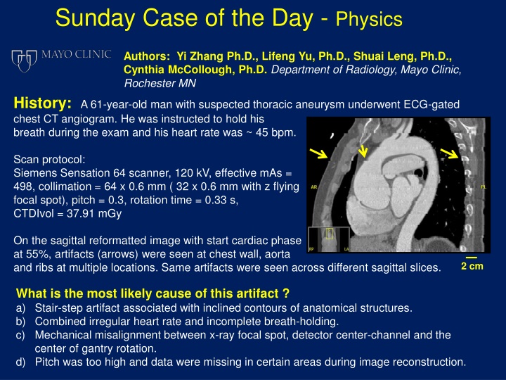
Sunday Case of the Day - Physics Authors: Yi Zhang Ph.D., Lifeng Yu, Ph.D., Shuai Leng, Ph.D., Cynthia McCollough, Ph.D. Department of Radiology, Mayo Clinic, Rochester MN History:A 61-year-old man with suspected thoracic aneurysm underwent ECG-gated chest CT angiogram. He was instructed to hold his breath during the exam and his heart rate was ~ 45 bpm. Scan protocol: Siemens Sensation 64 scanner, 120 kV, effective mAs = 498, collimation = 64 x 0.6 mm ( 32 x 0.6 mm with z flying focal spot), pitch = 0.3, rotation time = 0.33 s, CTDIvol = 37.91 mGy On the sagittal reformatted image with start cardiac phase at 55%, artifacts (arrows) were seen at chest wall, aorta and ribs at multiple locations. Same artifacts were seen across different sagittal slices. 2 cm What is the most likely cause of this artifact ? a) Stair-step artifact associated with inclined contours of anatomical structures. b) Combined irregular heart rate and incomplete breath-holding. c) Mechanical misalignment between x-ray focal spot, detector center-channel and the center of gantry rotation. d) Pitch was too high and data were missing in certain areas during image reconstruction.
Diagnosis: Correct answer is (d). Pitch was too high and data were missing in certain areas during image reconstruction. Retrospectively angiogram requires image reconstructed at the proper cardiac phase and at the proper z location. With high pitch and low heart rate, the table travels beyond the desired z-locations at the pre-determined cardiac phases, causing gaps between successive axial slices. Large gaps cannot be completely corrected by interpolation and cause discontinuities and banding artifacts in reformatted images. ECG-gated CT
Discussion Retrospectively ECG-gated CT angiogram requires image reconstructed at the proper cardiac phase and at the proper z location. To minimize the cardiac motion, only data acquired at the same cardiac phases are used. Also, the acquisition window is limited to a small fraction of the cardiac cycle. As illustrated in the figure below, reconstructions (yellow boxes) start at 55% of the R- R interval and last for ????= 0.33?. The maximal z-direction coverage is determined by the total collimator width: ?? = 32 0.6 = 19.2 ?? . With helical pitch 0.3, the patient table is moving at ??=?? ???? complete cardiac cycle ???= 23.3 ??. ???. After a = 17.5 ???? 60 ? 45 ???= 1.33 ?, the patient is move by ??= ?? ???= Because ??> ??, there are periodic gaps in z-direction where data were not acquired. Although small gaps can be interpolated acquisitions, larger gaps lead to discontinuities and banding artifacts in the reformatted images. by adjacent For ? ??, the pitch is bounded by: ???? ???? ???
Discussion ???? ??? , to compensate for 1) the In practice, the helical pitch is selected to be smaller than heart rate variation during the scan, and 2) decreased volume coverage due to helical motion and cone-beam geometry(1). (a), stair-step artifact associated with inclined contour can be seen in the reformatted image for helical scan, is incorrect. When the high contrast object is sloped along z- direction, the contour positions are different for reconstruction that takes measurement from one detector row to that which takes interpolations from two detector rows. This position difference is periodic, causing groove on the object contour along z-direction. However, this is not the case here, because the magnitude of this stair-step artifact is directly proportional to the table increment (TI) for low and moderate pitches(2). For our study here with TI about 0.5 cm, this artifact is almost invisible. (b) is incorrect because although cardiac or respiratory motion can cause phase mis- registration that appears as a banding artifact(3), it s usually more severe at the heart than that at the chest wall. (c) is incorrect. The misalignment between x-ray focal spot, detector center channel and the center of rotation causes the measured locations of projection rays to deviate from the ray positions assumed by the filtered back-projection, which results in image smearing. Combined with asymmetric helical weighting scheme, the smearing direction is periodic and can be seen as a zig-zag pattern on the high contrast object contour in the reformatted image(4). The bandings here are across the entire image rather than restricted to the anatomical structure boundaries.
References 1) Hsieh, J. (2009). Computed tomography: principles, design, artifacts, and recent advances. Bellingham, WA, SPIE. 2) Fleischmann, D., G. D. Rubin, et al. (2000). "Stair-Step Artifacts with Single versus Multiple Detector-Row Helical CT1." Radiology 216(1): 185-196. 3) Nakanishi, T., Y. Kayashima, et al. (2005). "Pitfalls in 16 Detector Row CT of the Coronary Arteries." Radiographics 25(2): 425-438. 4) Hsieh, J. (1999). Investigation of 3D image artifact caused by projection weighting and misalignment. Image Processing, 1999. ICIP 99. Proceedings.
