Assessment of a Newborn at 33 Weeks Gestation
Attending the assessment of a newborn at 33 weeks gestation, born to a mother with minimal prenatal care. The infant presents with mild jaundice, petechiae, and other concerning symptoms indicating a possible congenital infection. Lab results show anemia, thrombocytopenia, and elevated liver enzymes. Subsequent development of seizures and cranial CT findings further support the suspicion of congenital infection. Ophthalmological evaluation for chorioretinitis is recommended.

Download Presentation

Please find below an Image/Link to download the presentation.
The content on the website is provided AS IS for your information and personal use only. It may not be sold, licensed, or shared on other websites without obtaining consent from the author.If you encounter any issues during the download, it is possible that the publisher has removed the file from their server.
You are allowed to download the files provided on this website for personal or commercial use, subject to the condition that they are used lawfully. All files are the property of their respective owners.
The content on the website is provided AS IS for your information and personal use only. It may not be sold, licensed, or shared on other websites without obtaining consent from the author.
E N D
Presentation Transcript
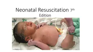
CASE SCENARIO: You are asked to attend assessment of a newborn of a 33-week gestation whose estimated birth weight is 1800 g. The mother is a 26-year-old G5,P4+0 who was admitted in active labour 2hours ago. The mother did not seek any prenatal care during pregnancy.
CASE SCENARIO: She reports no medical problems during the pregnancy. She denies tobacco, alcohol and illicit drug use. Mother's blood type is O+, antibody negative. Membranes are ruptured at the time of delivery revealing clear amniotic fluid.
CASE SCENARIO: At delivery, the baby is small male who was brought to the warming table. He is pink with good respiratory effort and his heart rate is 150 beats per minute. But, You note his skin is mildly jaundiced with raised red/purple lesions. So, you inform the mother that because you suspect the infant has a congenitally acquired infection, you are transferring him to the special care nursery for a more detailed examination and further management.
CASE SCENARIO: P/E of Newborn: Vital signs: Temperature: 37.5 C. Pulse: 120 bpm. RR: 40 breath/min. BP: 60/36 mmHg. O2 saturation: 100% in room air. Growth parameters: Weight: 1854 g. Length: 44 cm. Head circumference: 31.5 cm.
CASE SCENARIO: P/E of Newborn: He is a small, thin male infant. Has little subcutaneous fat. He is in no acute distress. Skin is mildly jaundiced with the "blueberry muffin" appearance of diffuse raised red/purple lesions and petechiae. His anterior fontanelle is soft, but full. Abdominal Exam: Distended. A firm liver edge is felt 4 cm below the right costal margin and the spleen is felt 3 cm below the left costal margin.
CASE SCENARIO: Lab.: CBC: Moderate anemia. Thrombocytopenia. Lymphocytosis. LFTs: Liver enzymes are elevated. Direct bilirubin: 8mg/100ml Elevated .
CASE SCENARIO: Three hours after birth, the infant develops generalized tonic-clonic seizures that stop after administration of 20mg/kg of phenobarbital. Cranial CT: Periventricularcalcification.
CASE SCENARIO: You suspected congenital infection with: CMV So, you consulted ophthalmologist to evaluate the patient for chorioretinitis.
Congenital Infections: Infections acquired in utero or during the birth process. They are a significant cause of fetal and neonatal mortality. Also, they are an important contributor to congenital malformation.
Congenital Infections: The concept of the perinatal infections is to group five infections in an acronym TORCH . T: Toxoplasmosis. O: Other; e.g., syphilis, parvovirus B19, R: Rubella. C: CMV. H: Herpes simplex virus HSV. These infections have similar presentations, including rash and ocular findings.
Index of Suspicion When do you think of TORCH infections? IUGR infants. HSM. Thrombocytopenia. Unusual rash. Concerning maternal history. Classic findings of any specific infection.
Diagnosing TORCH Infection Good maternal/prenatal history: Remember most infections of concern are mild illnesses often unrecognized. Thorough exam of infant. Directed labs/studies based on most likely diagnosis.
Toxoplasma gondii is a protozoan Organism exists in three forms Trophozoite Cyst Oocyst The incidance of congenetal toxplasma is0.3 to 1 /1000 live birth
What is the cause of toxoplasmosis? There are only a few ways to acquire the parasite that causes toxoplasmosis: Contact with cats or cat feces Eating raw or undercooked meat Drinking raw milk from an infected goat In 1sttrimester 17% spontaneous abortion 2ndtrimester 25% spontaneous abortion or sever disease 3rdtrimester 65% >>>>>subclinical disease
TOXOPLASMOSIS EPIDEMIOLOGY
Most infections are asymptomatic((70%)) When symptoms are present, they are will be ocular(76%) more than CNS manifestaion(52%) The ocular manifestation is chorioretinitis,optic atrophy, microphthalmia>>>>>blindness((use fundoscope))
CNS manefitation is hydrocephlus, intellctual and mental retardition, SNHL, seizure The triad of toxoplasmosis is Hydrocephalus Chorioretinitis Intracranial calcification>>do CT scan
TOXOPLASMOSIS TOXOPLASMOSIS DIAGNOSIS DIAGNOSIS Do cordocentisi and plcental sample Serology Toxoplasma IgG antibody IgM fluoresentantibody
Treatment of mother while fetus is still in utero Early treatment of the infant Compination of sulfadizin , pyramethamin, folic acid In pregnancy give her spyramycin
PREVENTION OF CONGENITAL PREVENTION OF CONGENITAL TOXOPLASMOSIS TOXOPLASMOSIS Use precautions when handling cat litter box Do not eat inadequately cooked meat
CONGENITAL INFECTIONS CONGENITAL INFECTIONS CONCLUSIONS CONCLUSIONS Congenital toxoplasmosis key is avoidance of exposure during pregnancy
CONGENITAL RUBELLA caused by an RNA Togavirus . ssRNA virus. Vaccine-preventable disease . Transmission: To mother: respiratory droplets. To fetus: Transplacental. The greatest risk for transmission to the fetus is during the 1sttrimester.
Rubella: Rubella: . Neonatal Manifestations IUGR low birth weight - prematurity stillbirth - spontaneous abortion Early Manifestations cloudy corneas Cataracts microcephaly hepatomegalysplenomegaly jaundice pulmonary valve stenosis patent ductus arteriosus thrombocytopenia purpura
MANIFESTATIONS OF CONGENITAL RUBELLA 80 70 60 50 40 30 20 10 0 Deafness Eye CNS Cardiac
PDA Sensorineural Hearing Loss Cataract
Effect on the Mother: Rash. Fever. Lymphadenopathy. Arthralgia.
Diagnosis: Virology Can isolate virus from nasal secretions Less frequently from throat, blood, urine, CSF Serology fetal rubella-specific IgM persistence of rubella-specific IgG after 8-12 months of age
Treatment: No specific treatment. Avoid rubella vaccine during pregnancy
Congenital CMV infection Defined as the isolation of CMV from the saliva or urine within 3 weeks of birth. member of the herpesvirus [ DNA ] Most common congenital viral infection Leading cause of sensorineural deafness Major cause of mental retardation, cerebral palsy CMV doesnotaffect organogenesis but affects organ already developed Approximately 0.15 2% of live births
How is CMV transmitted? - Fetus: Via placenta from the mother - Human milk - Blood transfusion, organ transplantation - Children and adults: Mainly via bodily fluids (esp. urine, saliva) - For pregnant women, the two most common exposures to CMV are through sexual contact and through contact with the urine and saliva of young children with CMV infection.
CMV CLINICAL MANIFESTATIONS CNS Manifestation : 70% microcephaly 60% intellctual impairment 35% sensorineural hearing loss 7% sizure
CMV CLINICAL MANIFESTATIONS Systemic manifestation: Hepatosplenomegaly (70%) Jaundice (68%) Thrombocytopenia with Petechiae Chorioretinitis (20%) ) ( 65%
SEQUELAE OF SYMPTOMATIC CONGENITAL CMV INFECTION Seizures Chorioretinitis Periventricularcalcifications Sensorineural hearing loss motor deficits
CHARACTERISTICS ASSOCIATED WITH INCREASED RISK OF SEQUELAE Primary maternal infection Symptomatic congenital CMV infection Presence of neonatal neurological abnormalities Abnormal head CT scan Chorioretinitis in the newborn
Diagnosis of Congenital CMV Infections Isolation of CMV from urine or other body fluid (,CSF, blood, saliva) in the first 21 days of life. [gold standard] Serologic tests; CMV-specific IgM. (false +ve , false ve ) PCR
Evaluation of mothers at risk of transmitting CMV to the fetus Test for IgG antibody at first prenatal visit Positive Negative Test for IgM Antibody Retest later Negative, no further testing Positive = primary infection IgG Positive = Seroconversion Negative, no further tests Refer for prenatal diagnosis
Treatment for Babies Born with CMV If your baby is diagnosed with congenital CMV infection, you should have his or her hearing and vision checked regularly. There is some evidence that ganciclovir, an antiviral drug, may prevent hearing loss and developmental outcomes in infants born with symptomaticcongenital CMV infection with central nervous system involvement.
How is congenital CMV prevented? Here are a few simple steps you can take to avoid exposure to saliva and urine that might contain CMV : Wash your hands often with soap and water for especially after changing diapers feeding a young child wiping a young child s nose or drool handling children s toys Do not share food, drinks, or eating utensils used by young children Do not put a child s pacifier in your mouth Do not share a toothbrush with a young child Avoid contact with saliva when kissing a child Clean toys, countertops, and other surfaces that come into contact with children s urine or saliva 20 15 - seconds,


)")