Assessment of Coagulation Profile and Thrombocyte Levels in Sudanese Neonates with Sepsis
This study conducted at Omdurman Maternity Hospital in Sudan aimed to evaluate the impact of sepsis on coagulation profile and thrombocyte levels in neonates. The research assessed platelet counts, prothrombin time, fibrinogen levels, and various other factors in septic neonates compared to healthy controls. Early detection of sepsis in neonates is crucial for prompt treatment with appropriate antimicrobial medications.
Download Presentation

Please find below an Image/Link to download the presentation.
The content on the website is provided AS IS for your information and personal use only. It may not be sold, licensed, or shared on other websites without obtaining consent from the author. If you encounter any issues during the download, it is possible that the publisher has removed the file from their server.
You are allowed to download the files provided on this website for personal or commercial use, subject to the condition that they are used lawfully. All files are the property of their respective owners.
The content on the website is provided AS IS for your information and personal use only. It may not be sold, licensed, or shared on other websites without obtaining consent from the author.
E N D
Presentation Transcript
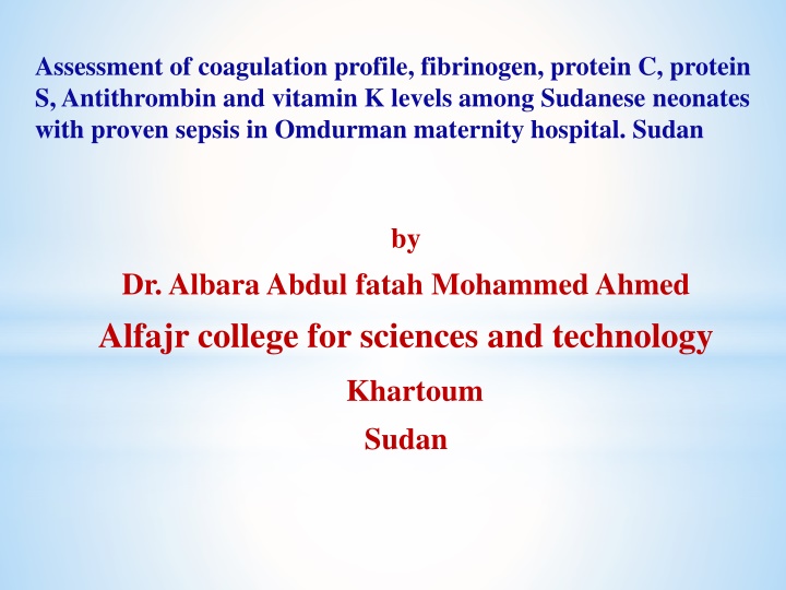
Assessment of coagulation profile, fibrinogen, protein C, protein S, Antithrombin and vitamin K levels among Sudanese neonates with proven sepsis in Omdurman maternity hospital. Sudan by Dr. AlbaraAbdul fatah Mohammed Ahmed Alfajr college for sciences and technology Khartoum Sudan
Background The neonatal sepsisincreases the cost of medical care, as it causes severe morbidities and therefore increases the length of hospitalization in neonatal intensive care units (NICU) (1, 2). Thus neonatal sepsis accounts over 50% of the neonatal deaths (3, 4). Early-onset sepsis among neonates is caused by organisms in the birth canal either when the amniotic membranes rupture or leak prior or during the delivery (5). In contrast, late-onset sepsis in neonates might be acquired from the environment (1). Previous studies reported that, medical laboratory scientists and clinicians have faced challenges to recognize the neonatal septic (6). Accordingly, septic neonates must be diagnosed rapidly for taking the right antimicrobial medications (6,3,7).
Moreover, some laboratory investigations should be done such as blood culture and platelet count to alleviate the medical complications of this illness (3, 4). Accordingly, the current study aimed to detect the effect of sepsis on haemostatic tests and circulating thrombocytes among Sudanese neonates with sepsis.
Objectives : The study aimed to assess the platelet counts, prothrombin time (PT), activated partial thromboplastin time (APTT), thrombin time (TT), fibrinogen, Protein C, protein S, antithrombin (AT) , and vitamin K in Sudanese neonates with sepsis and compare them with healthy neonates in order to study haemostatic alteration among septic neonates.
Methods and patients This study was a prospective case control study. It was conducted in Omdurman maternity hospital in the period from June 2013 to April 2015. Populations: Figures 1 and 2 define the populations in this study. Figure 1A describes the patient population of 50 septic neonates, 26 female and 24 male individuals. Figure 1B defines a control group comprising 27 females and 24 males, numbers expressed as percentages on the graphics.
Study area: The study was conducted in Omdurman maternity hospital from June 2013 to April 2015 comprising 50 consecutive sepsis cases and 50 controls. Bacterial culture: Blood culture, Gram stain, culture, sensitivity and biochemical tests were done for all neonates with suspected sepsis for bacterial identification and diagnosis neonates with sepsis. Platelet count: EDTA microtainer was used for platelets count, using cell counter, Sysmex KX-21 analyzer). Haemostatic studies: Platelet poor plasma collected from trisodium citrate was used for haemostatic studies, PT, APTT, TT, fibrinogen, proteins C and S, using a Stago coagulometer. Antithrombin (AT) was assessed by turbidimetrically (Mindray BA-88 Vitamin K was assessed by high performance liquid chromatography (HPLC), Schmiatzu. 10 ADVP after samples were centrifuged, precipitated, extracted, evaporated and injected into HPLC. Data analysis:- Data was analyzed by t-test using SPSS 20. ).
Result Delivery mode Figure 2A & B Mode of delivery among patient groups Patient and control groups were further classified into caesarean section and normal vaginal delivery neonates. Of the patient group 17 neonates were delivered normally and 33 neonates were delivered by caesarean section (Figure 2A). In the control group 7 neonates were delivered normally, 43 were delivered by caesarean section as shown in Figure 2B. Proportions are given as percentages on the graphics.
Figure 3A & B Gestational age among study population According to the gestational age, the patient and control groups were classified into term and preterm neonates. Of the patient group, was 17 neonates (34%) were term and 33 neonates (66%) were preterm (Figure 3A). Of the control group, only 2% (one neonate) was preterm and 98% (49 neonates) were term, as shown in (Figure 3B).
Parameter Platelets count (test) Platelets count (control) Mean 60,289 c/mm3 212,030 c/mm3 Std.Deviation 52,494 62,092 P.value 0.000 PT (test) PT (control) 16.6 sec 13.9 sec 7.61 1.97 0.018 APTT (test) APTT (control) 47.8 sec 37.5 sec 16.2 5.06 0.000 TT (test) TT (control) 18.7 sec 20.5 sec 3.81 3.10 0.009 Fibrinogen (test) Fibrinogen (control) 482.2 mg/dl 393.7 mg/dl 180.7 103.2 0.003 Protein C (test) Protein C (control) 34.3 % 36.8 % 15.8 12.9 0.412 Protein S (test) Protein S (control) 33.4 % 34.7 % 20.8 20.9 0.762 Antithrombin (test) Antithrombin (control) 183.9 Mg/ml 221.5 Mg/ml 69.6 71.9 0.003 Vitamin K (test) Vitamin K (control) 0.86 ng/ml 1.23 ng/ml 1.10 1.11 0.105
Causative bacterial and its mortality in the patient group. Table 1 demonstrates eight types of bacteria were isolated among septic neonates, Pseudomonas, Staph.epidermidis, Strep. fecailis, E.coli, Staph.aureus, non groupB Streptococci. The Table also shows frequency of mortality which was caused by Pseudomonas), Salmonella, S. Epidermidis, Klebsiella, and E. coli. No mortality occurred in patients with Strep. fecailis, Staph. aureus and Streptococci (Non group B). Salmonella, Klebsiella,
Table 1 demonstrates Causative bacterial and its mortality in the patient group. Key: Significance determined by comparison of each group with controls indices. (a) =P<0.01; (b) = p < 0.05; (c) = p >0.05.*Statistical significance shown in parenthesis.
Haemostatic tests according to the outcome of the patient group The Protein C and vitamin K were significantly lower in the dead patients than in the recovered patients(p< 0.05), whereas APTT was significantly higher in the dead patients, as shown in Table 2. In contrast, the following parameters were significantly lower in patients (case group) than in control samples: PT, APTT and fibrinogen (p<0.001). The difference in platelet count, PT, TT, protein S and antithrombin was not significant in the dead patients with the recovered patients (See Table 2 ).
Table 2 shows haemostatic tests according to the outcome of the patient group Key: Significance determined by comparison of each group with controls indices. (a) =P<0.01; (b) = p < 0.05; (c) = p >0.05.*Statistical significance shown in parenthesis.
Figure 4 shows onset of sepsis among patient group. According to the onset of sepsis, the mortality in the patient group (ten patients in total) were classified intoearly(from birth-7 days) and late onset sepsis(8-28 days). The former was 40% (N=4) and late onset sepsis 60% (N=6).
Protein C and vitamin K according to the outcome of the patient group Table shows haemostatic profile based on the early onset of the patient group. The Protein C and vitamin K were significantly lower in the late onset patients than in the recovered patients(p< 0.05), whereas APTT was significantly higher in the dead patients In contrary, the following parameters were significantly lower in patients than in control samples: PT, APTT and fibrinogen (p<0.001). The difference in platelet count, PT, TT, protein S and antithrombin was not significant in the dead patients with the recovered patients.
Table 4. Haemostatic tests according to the onset of sepsis of the patient group
Table 5. Haemostatic tests according to the Gram stain types in case group
Table 6. Results of coagulation profile in the patient group stratified by causative bacteria 54.7
Conclusion and recommendations It has been concluded that platelets count significantly decreased, both PTandAPTTsignificantly prolonged. TTsignificantly shorted, fibrinogen was significantly increased andAT significantly decreased in neonatal sepsis. APTT and protein C showed significant correlation with the outcome, so both can predict early mortality, PTandTTshowed significant correlation with early onset sepsis. Demographic data (gender, gestational age, mode of delivery, and Gram stain typing had no effecton haemostatic parameters.
Recommendations 1-Coagulation assessment (including platelets count, PT, APTT, TT, fibrinogen) should be involved in the criteria of evaluation of neonatal sepsis. 2-The APTT and protein C can be used to predict mortality among septic neonates. PT and TT predicts the onset of sepsis. 3- More prospective intervention studies can be applied on vitamin k to study its effect on progression of neonatal sepsis and focus on its outcome. 4- Preventive measures can be taken to minimize the occurrence of neonatal sepsis, and special care for those of primigravida. 5- More studies on neonatal sepsis are needed
References 1.Kung YH, Hsieh YF, Weng YH, Lien RI, Luo J, Wang Y, et al. Risk factors of late-onset neonatal sepsis in Taiwan: A matched case-control study. J Microbiol Immunol Infect. 2016 Jun;49(3):430-5. 2.Tzialla C, Borghesi A, Serra G, Stronati M, Corsello G. Antimicrobial therapy in neonatal intensive care unit. Ital J Pediatr. 2015 Apr 01;41:27. 3.Chiesa C, Panero A, Osborn JF, Simonetti AF, Pacifico L. Diagnosis of neonatal sepsis: a clinical and laboratory challenge. Clin Chem. 2004 Feb;50(2):279-87 Stefanovic IM. Neonatal sepsis. Biochem Med (Zagreb). 2011;21(3):276-81. 4-Kara S, Emeksiz Z, Alioglu B, Dallar Bilge Y. Effects of neonatal sepsis on thrombocyte tests. J Matern Fetal Neonatal Med. 2016;29(9):1406-8 5- Ofman G, Vasco N, Cantey JB. Risk of Early-Onset Sepsis following Preterm, Prolonged Rupture of Membranes with or without Chorioamnionitis. Am J Perinatol. 2016 Mar;33(4):339-42. 6.Simonsen KA, Anderson-Berry AL, Delair SF, Davies HD. Early-onset neonatal sepsis. Clin Microbiol Rev. 2014 Jan;27(1):21-47. 7.De Assis Meireles L, Vieira AA, Costa CR. [Evaluation of the neonatal sepsis diagnosis: use of clinical and laboratory parameters as diagnosis factors]. Rev Esc Enferm USP. 2011 Mar;45(1):33-9.


