Atlanto-Occipital Dissociation: Traumatic Mechanism and Imaging Findings

Atlanto-Occipital Dissociation (AOD) is a severe neck injury resulting from high-velocity trauma. This condition has a high mortality rate, with key imaging findings including basion-dens interval, atlanto-occipital interval, and condylar sum measurements on CT scans. Ligamentous disruption at the craniocervical junction determines the stability of AOD, with the tectorial membrane and alar ligaments being crucial stabilizers. Differential diagnosis includes atlantoaxial distraction and rotatory subluxation. MRI is essential for confirming the diagnosis and guiding treatment decisions.

Download Presentation

Please find below an Image/Link to download the presentation.
The content on the website is provided AS IS for your information and personal use only. It may not be sold, licensed, or shared on other websites without obtaining consent from the author.If you encounter any issues during the download, it is possible that the publisher has removed the file from their server.
You are allowed to download the files provided on this website for personal or commercial use, subject to the condition that they are used lawfully. All files are the property of their respective owners.
The content on the website is provided AS IS for your information and personal use only. It may not be sold, licensed, or shared on other websites without obtaining consent from the author.
E N D
Presentation Transcript
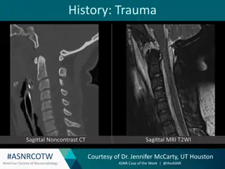
History: Trauma #ASNRCOTW American Society of Neuroradiology Courtesy of Dr. Jennifer McCarty, UT Houston ASNR Case of the Week | @theASNR
Atlanto-Occipital Dissociation (AOD) Mechanism: High velocity trauma w/ distraction type injury Mortality: A reported 90% of patients w/ AOD injuries die, 60% at the scene. Three Types: I Ventral dislocation, most common II Longitudinal distraction, most unstable III Dorsal dislocation Imaging Findings on CT Basion-dens interval > 10 mm in adults Atlanto-occipital interval > 2 mm Condylar Sum > 4.2 mm, most sensitive CT sign Ligamentous disruption may involve any of the craniocervical junction ligaments, but the most important stabilizers are: Tectorial membrane Alar ligaments #ASNRCOTW American Society of Neuroradiology Courtesy of Dr. Jennifer McCarty, UT Houston ASNR Case of the Week | @theASNR
Additional Info Differential diagnosis: Atlantoaxial Distraction Multiple choice question What is the diagnosis? Atlanto-Occipital Dissociation Occipital Condylar Fracture Atlantoaxial Distraction Rotatory Subluxation Reference link: https://pubs.rsna.org/doi/10.1148/rg.2015150035 Diagnosis confirmed with MRI #ASNRCOTW American Society of Neuroradiology Courtesy of Dr. Jennifer McCarty, UT Houston ASNR Case of the Week | @theASNR
Slide Example Gradient: Standard Large Arrows (Light): Standard Small Arrows (Light): Standard Large Arrows (Dark): Standard Small Arrows (Dark): #ASNRCOTW American Society of Neuroradiology Courtesy of ASNR Case of the Week | @theASNR

")