Autonomic Nervous System: Overview and Components
The autonomic nervous system (ANS) controls involuntary body functions through its sympathetic and parasympathetic branches. It consists of visceral receptors, sensory neurons, control centers in the CNS, preganglionic neurons, ganglia, postganglionic neurons, and effectors. Neurotransmitters like acetylcholine and norepinephrine play key roles. Receptors in the ANS bind with these neurotransmitters, influencing various bodily responses. Explore the intricate workings of the ANS for a deeper insight into its regulatory functions.

Download Presentation

Please find below an Image/Link to download the presentation.
The content on the website is provided AS IS for your information and personal use only. It may not be sold, licensed, or shared on other websites without obtaining consent from the author.If you encounter any issues during the download, it is possible that the publisher has removed the file from their server.
You are allowed to download the files provided on this website for personal or commercial use, subject to the condition that they are used lawfully. All files are the property of their respective owners.
The content on the website is provided AS IS for your information and personal use only. It may not be sold, licensed, or shared on other websites without obtaining consent from the author.
E N D
Presentation Transcript
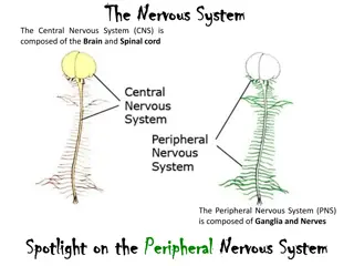
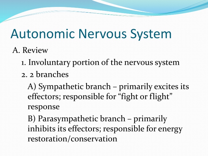
Autonomic Nervous System A. Review 1. Involuntary portion of the nervous system 2. 2 branches A) Sympathetic branch primarily excites its effectors; responsible for fight or flight response B) Parasympathetic branch primarily inhibits its effectors; responsible for energy restoration/conservation
Autonomic Nervous System A. Components of the ANS 1. Visceral receptors 2. Visceral sensory neurons 3. Autonomic control centers in CNS 4. Preganglionic (motor) neurons (Sympathetic & Parasympathetic)
Autonomic Nervous System 5. Autonomic ganglia (S&P) 6. Postganglionic (motor) neurons (S&P) 7. Autonomic effectors (S&P; often a single effector is shared by both branches)
Autonomic Nervous System B. Neurotransmitters 1. Two neurotransmitter are released by the motor neurons of the ANS (components #4 & 6) A) Acetylcholine (ACh) released from cholinergic neurons 1) All sympathetic and parasympathetic preganglionic neurons release ACh
Autonomic Nervous System 2) All parasympathetic postganglionic neurons release ACh 3) A few sympathetic postganglionic neurons (ex. sweat glands) release ACh B) Norepinephrine released from adrenergic neurons 1) Most sympathetic postganglionic neurons release norepinephrine
Autonomic Nervous System C. Receptors are found on the dendrites of the postganglionic neurons in the autonomic ganglia, and on the autonomic effectors (Refer to the summary table found on the lecture materials page) 1. Cholinergic receptors bind with ACh A) 2 types
Autonomic Nervous System 1) Nicotinic a) 17 different types, divided into 2 subtypes (neuronal & muscle) b) Found on the dendrites of all parasympathetic and sympathetic postganglionic neurons; also found on the adrenal glands (excitatory)
Autonomic Nervous System 2) Muscarinic a) 5 subtypes (M1-M5) b) Found throughout the body on all parasympathetic effectors i) Mostly excitatory but some are inhibitory dependent on location c) Found on a few sympathetic effectors sweat glands (excitatory)
Autonomic Nervous System 2. Adrenergic receptors bind with norepinephrine A) 2 types 1) Alpha ( ) adrenergic 2 subtypes a) 1 located on blood vessels supplying the skin, mucosa, viscera, and kidneys; also found on nearly all sympathetic effectors except the heart (excitatory) b) 2 located on the pancreas (inhibitory) and blood platelets (excitatory)
Autonomic Nervous System 2) Beta ( ) adrenergic 3 subtypes a) 1 located primarily on cardiac muscle tissue (excitatory) b) 2 located on blood vessels of heart, liver, and skeletal muscle tissue (inhibitory); also found on the lungs and most sympathetic effectors (mostly inhibitory) c) 3 located on adipose tissue (excitatory)
Autonomic Nervous System D. Comparison of Sympathetic & Parasympathetic 1. Origin of their preganglionic neurons A) Sympathetic originate from the thoracic & lumbar regions B) Parasympathetic originate from the brain & sacral regions 2. Location of their ganglia A) Sympathetic located near the spinal cord 1) Compose the sympathetic chain ganglia B) Parasympathetic located within the effector
Autonomic Nervous System 3. Length of their neurons A) Sympathetic short preganglionic neurons & long postganglionic neurons B) Parasympathetic long preganglionic neurons & short postganglionic neurons E. Parasympathetic Nervous System 1. Responsible for energy restoration & conservation 2. Cranial Outflow
Autonomic Nervous System A) Occulomotor Nerve (III) 1) Cause pupils to constrict and the lens to bulge (focusing on close objects) B) Facial Nerve (VII) 1) Stimulates large glands of the head (salivary, lacrimal, and nasal) C) Glossopharyngeal Nerve (IX) 1) Stimulates parotid salivary gland
Autonomic Nervous System D) Vagus Nerve (X) 1) Decreases/steadies heart rate and constricts coronary vessels 2) Constricts bronchioles 3) Expels bile from gallbladder 4) Stimulates secretion of enzymes in the stomach 5) Increases motility (peristalsis) and relaxes the sphincter muscles in the intestines
Autonomic Nervous System 3. Sacral Outflow A) Relaxes internal anal sphincter to promote defecation B) Contraction of smooth muscle of urinary bladder wall and relaxes internal urethral sphincter to promote urination C) Causes dilation of the vessels leading to the genitalia causing penile or clitoral erection
Autonomic Nervous System F. Sympathetic Nervous System 1. Responsible for the fight-or-flight response 2. 2 groups of sympathetic ganglia A) Sympathetic chain ganglia 1) Lie in a vertical row on either side of the vertebral column
Autonomic Nervous System 2) Generally innervate organs above the diaphragm 3) Examples: a) Superior Cervical Ganglia i) Innervates structures of the head including sweat & salivary glands and smooth muscles of the eye
Autonomic Nervous System b) Middle & Inferior Cervical Ganglia i) Innervate structures of the thorax including the heart and thyroid gland B) Collateral (prevertebral) ganglia 1) Lie anterior to the spinal cord and close to the large abdominal arteries 2) Generally innervate structures below the diaphragm
Autonomic Nervous System 3) Examples: a) Celiac Ganglia i) Decreases digestive and urinary functions ii) Stimulates release of glucose from liver and the release of adrenal hormones b) Superior Mesenteric Ganglia i) Decreases small intestine activity
Autonomic Nervous System c) Inferior Mesenteric Ganglia i) Decreases large intestine activity d) Hypogastric Ganglia i) Relaxes the walls of the urinary bladder and constricts the internal urethral sphincter to inhibit urination ii) Causes ejaculation or vaginal contractions
Hypogastric Ganglion
Autonomic Nervous System G. Summary 1. Sympathetic NS A) Increases heart rate & blood pressure B) Increases respiration (rate & depth) C) Increases blood glucose levels D) Decreases the activities of the digestive and urinary systems
Autonomic Nervous System 2. Parasympathetic NS A) Decreases heart rate B) Decreases blood pressure (both directly & indirectly) C) Decreases respiration (rate & depth) D) Decreases blood glucose levels E) Increases digestive function F) Has no direct effect on kidney function