Avian Trichomoniasis: Symptoms, Transmission, and Treatment
Avian trichomoniasis is a parasitic disease affecting young birds and caused by Trichomonas gallinae. It can lead to sudden death in acute cases and is transmitted through carriers, contaminated water and feed, and wild birds. Symptoms include nodules in the oral cavity, weight loss, ruffled appearance, and diarrhea. Diagnose through microscopic examination and treat with antiprotozoal medications. Proper management includes culling carrier birds and maintaining clean food and water sources to prevent reinfection.
Download Presentation

Please find below an Image/Link to download the presentation.
The content on the website is provided AS IS for your information and personal use only. It may not be sold, licensed, or shared on other websites without obtaining consent from the author.If you encounter any issues during the download, it is possible that the publisher has removed the file from their server.
You are allowed to download the files provided on this website for personal or commercial use, subject to the condition that they are used lawfully. All files are the property of their respective owners.
The content on the website is provided AS IS for your information and personal use only. It may not be sold, licensed, or shared on other websites without obtaining consent from the author.
E N D
Presentation Transcript
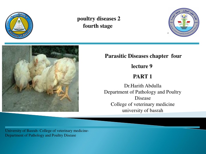
poultry diseases 2 fourth stage Parasitic Diseases chapter four lecture 9 PART 1 Dr.HarithAbdulla Department of Pathology and Poultry Disease College of veterinary medicine university of basrah University of Basrah- College of veterinary medicine- Department of Pathology and Poultry Disease
Avian trichomoniasis is a disease of young birds. Adult birds that recover from the infection may still carry the parasite, but are resistant to reinfection. Trichomonas gallinae is a common parasite of pigeons and doves. Other birds such as domestic and wild turkeys, chickens may also become infected.
Trichomonas oral-nasal cavity or anterior end of the digestive and respiratory tracts. Trichomonas gallinae gallinae is generally found in the Transmission: 1-From bird to bird through: a a- -Carriers. b b- -Contaminated water and feed. c c- -Wild pigeons and other birds. Transmission:
In acute cases (Adults): indication that the bird is infected, and death may occur quite suddenly. In acute cases (Adults): There may be little In other cases(Young): nodules in the oral cavity, esophagus and crop strongly suggest trichomoniasis. In other cases(Young): Characteristic yellowish-white Infected birds may stop feeding, lose weight, look ruffled and dull, and be unable to stand or maintain their balance. Diarrhea may also occur. Death may occur within three weeks of infection.
In young : 1-Small white to yellowish areas in the mouth cavity, especially the soft palate. In young : 2-Inflammation and ulceration of the mucosal surface. 3-Greenish fluid or cheesy material may accumulate in the mouth and crop, and this material may exude from the beak. 4- Large, firm necrotic masses that may block the lumen. 5-The disease may spread by penetrating the underlying tissues to involve the liver and other organs.
In adults: In adults: None. Diagnosis: 1-Microscopic examination of the greenish fluids, cheesy material . 2- The lesion. Diagnosis:
1-Culling or treating carrier birds. 2- Food and water sources should be cleaned regularly and protected from contamination by wild pigeons and other birds. Treatment: Antiprotozoal medications, such as dimetridazole and metronidazole. Treatment:
Histomoniasis birds, particularly chickens and turkeys, caused Histomoniasis (Blackhead,Enterohepatitis) is a disease of by a protozoan, Histomonas meleagridis. The parasite specifically infects the ceca and liver. Heterakis gallinarum (Cecal worm) Ingest Histomonas meleagridis Ingested by Earthworms, the latter taken by birds Infection.
Depression. Inappetance. Poor growth. Sulphur-yellow diarrhea. Cyanosis of head. Blood in faeces (chickens). Progressive depression and emaciation.
Enlargement of caeca. Ulcers, caseous cores with yellow, grey or green areas. Liver may have irregular-round depressed lesions, usually grey in colour, they may not be present in the early stages, especially in chickens.
1.Lesions 2.Scraping from fresh material for microscopic examination. Prevention: 1-Good sanitation, avoid mixing species and concrete floors. 2-Use of an anti-histomonas product in feed. Prevention:
1-Nitro-imidazoles (e.g. dimetridazole). 2-Nitrofurans (e.g. furazolidone, nifursol). 3-Arsenicals (e.g. nitarsone) .
Cryptosporidia are related to the coccidia, but much smaller (typically oocysts are less than of the size of an Eimeria acervulina oocyst). They also differ from coccidia in being poorly host specific. Cryptosporidium baileyi can cause respiratory disease in chickens and turkeys. The same species causes infections of the hindgut and cloacal bursa in chickens, turkeys, and ducks. Cryptosporidium meleagridis also infects both species. A further species causes respiratory disease in quail. The oocysts are excreted ready sporulated in the faeces and infection occurs by inhalation and ingestion.
. Cough. Swollen sinuses. Low weight gain. Diarrhea. Post Sinusitis. Airsacculitis. Pneumonia. Post- -mortem lesions: mortem lesions:
Microscopic examination: Identification of the parasites attached to the epithelium. Treatment: None If other disease processes are complicating (e.g. colisepticaemia) there may be benefit in medicating for these. Treatment: Prevention: The oocysts of cryptosporidia are extremely resistant to chemical disinfection. There are no effective preventative medicines or feed additives. Prevention: Steam cleaning is effective in reducing infection as oocysts are inactivated above about 65 C.
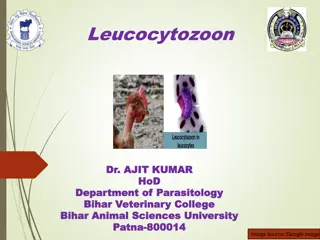
Infections with Leucocytozoon spp are most often subclinical but can occasionally be clinical and even fatal. Mortality may approach 100% in susceptible species and varies greatly with species and strain of parasite, host species, degree of exposure, and other factors.
1 1- -Acute: ( Young ). a-Listless. b- Anemia. c- Leukocytosis. d- Tachypnea. e-Anorexia. f-Diarrhea with green droppings. g-Often CNS signs. Acute: ( Young ). 2 2- -Subacute or ( Chronic): In Adults. a-Poor growth. b- Impaired egg production. Subacute or ( Chronic): In Adults. Visibly affected birds die after 7 10 days or may recover with sequelae of poor growth and egg production.
http://1.bp.blogspot.com/-hisYcCZ1Gbo/TwlJvHWHZ8I/AAAAAAAAADE/B9lnuhWByDk/s320/leu8.jpghttp://1.bp.blogspot.com/-hisYcCZ1Gbo/TwlJvHWHZ8I/AAAAAAAAADE/B9lnuhWByDk/s320/leu8.jpg
1-Hemorrhages. 2- Splenomegaly. 3-Hepatomegaly. 4-Grossly visible white dots in affected organs. Diagnosis: 1-Blood smear. 2-Serology may detect prior infection. Diagnosis:
1-Control invertebrate vectors are helpful. 2-Preventive medication using pyrimethamine and sulfadimethoxine combined in the feed. Treatments: Treatment usually is not effective. Quinacrine hydrochloride or trimethoprim /sulfamethoxazole solution have been used in raptors; the parasitemia is reduced but the infection is not cleared. Treatments:
Syngamous 1-Gape worm. 2-Red worm. 3-Forked worm. Syngamous trachea: trachea: Clinical signs: 1-The birds sneeze, cough, and frequently shake their heads in an effort to expel the worms and mucus from the windpipe. Clinical signs: 2- The affected birds stretch the neck and gape with open mouth for air.
In young birds, under 8 weeks of age, the activities of the worms cause inflammation of the trachea with accumulation of considerable amount of mucus. Prevention: *Frequent removal of litter to prevent accumulation of worm eggs and larvae. *Keep intermediate hosts out of the reach of chicken flocks. Prevention: Treatment: Treatment: Unsatisfactory.
a a- -Capillaria There are several species of Capillaria that occur in poultry. Capillaria annulata and Capillaria contorta occur in the crop and esophagus. Capillaria ( Capillary or thread worms). ( Capillary or thread worms). In the lower intestinal tract there may be several different species but usually Capillaria obsignata is the most prevalent.
1-Emaciation, weakness, bloody diarhea(Extreme). 2-Decrease egg production possible. Post Inflammation , thickening or hemorrhage of the gut wall. Post- -mortem lesions: mortem lesions: Diagnosis: 1-If present in large numbers, these parasites are usually easy to find at necropsy. 2-Fecal float- for presence of eggs. Diagnosis:
1-Proper management of diet 2-Sanitation. 3-Treatment. 4-Chickens need a proper diet, especially an adequate supply of vitamins A and B complex.
B B- - Ascarids One of the most common parasitic roundworms of poultry (Ascaridia galli) occurs in chickens and turkeys. Ascarids (Large Intestinal Roundworms). (Large Intestinal Roundworms). Of all the intestinal worms, it probably inflict the most damage. Young birds are affected more severely.
1-Unthriftyness. 2- Poor growth and feed conversion. 3-Decreased egg production. 4-Death in severe infections. Post 1-Mucosa damage from migrating larvae. 2-Hemorrhage. 3-Enteritis. Post- -mortem lesions: mortem lesions:
Macroscopic examination with identification of worms, oval smooth-shelled non- embryonated eggs in faeces. Control: Prevention of contamination of feeders and drinkers with faeces; pasture rotation and regular treatment, especially for young birds. Control: Treatment: Treatment: Piperazine is the drug of choice.
The parasite (Heterakis gallinae) is found in the ceca of chickens, turkeys and other birds. This parasite apparently does not seriously affect the health of the bird. At least no marked symptoms or lesions can be blamed on its presence. Its main importance is that it has been incriminated as a vector of Histomonas meleagridis, the agent that causes blackhead.
1-No marked signs. 2-Black head in turkeys as carrier of Histomonas meleagridis( Protozoan). Post 1-Thickening of cecal mucosa (lining). 2-Hemorrhages- only in heavy infection. Post- -mortem lesions: mortem lesions: Prevention together or allow turkeys on range which chickens have previously occupied. Prevention : Should never house chickens and turkeys Treatment: Treatment: Fenbendazole.
Cestodes are tapeworms that are seen in many species; they may not be host specific. Most have intermediate invertebrate hosts such as beetles or earthworms. Clinical signs: It is doubtful if any signs are produced under most circumstances. Clinical signs:
Occupy space in intestine and create small lesions at point of attachment. Diagnosis: Identification of the presence of the worms at post-mortem examination. Diagnosis:
Flubendazole. Prevention Control the intermediate hosts, or birds' access to them. Prevention
Clinical signs: 1-Lose their normal activity and appetite. 2-Dropping off in egg production. 3-The eggs that are produced frequently have very thin shells or no shells. Clinical signs: 1-Adhesive peritonitis might result. 2-Parasite may be found in oviduct or eggs. Treatment: Treatment: CC14
