Barrett's Esophagus: Metaplastic Change in Esophagus Lining
Barrett's esophagus is a metaplastic change in the lining of the esophagus due to chronic reflux. Regular surveillance endoscopy is crucial for early detection. Treatment focuses on managing underlying GERD to reduce the risk of cancer development.

Download Presentation

Please find below an Image/Link to download the presentation.
The content on the website is provided AS IS for your information and personal use only. It may not be sold, licensed, or shared on other websites without obtaining consent from the author.If you encounter any issues during the download, it is possible that the publisher has removed the file from their server.
You are allowed to download the files provided on this website for personal or commercial use, subject to the condition that they are used lawfully. All files are the property of their respective owners.
The content on the website is provided AS IS for your information and personal use only. It may not be sold, licensed, or shared on other websites without obtaining consent from the author.
E N D
Presentation Transcript
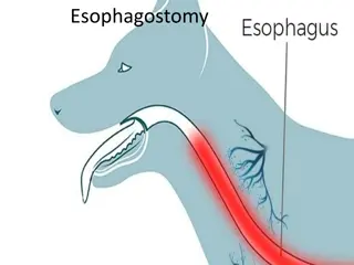
Oesophagus Lecture III Dr. Zaeem Fezea Dahla Consultant General Surgeon CABS, MD
LEARNING OBJECTIVES BENIGIN CONDITIONS OF ESOPHAGOUS
Barrett's Barrett'soesophagus oesophagus(columnar (columnar- -lined lined lower lower oesophagus oesophagus) ) Barrett's oesophagus is a metaplastic change in the lining mucosa of the oesophagus in response to chronic gastro- oesophageal reflux Many of these patients do not have particularly severe symptoms,although they do have the most abnormal pH profiles. One of the great mysteries of GORD is why some people develop oesophagitis and others develop Barrett's oesophagus,often without significant oesophagitis.
Barrett's oesophagus with proximal migration of the squamocolumnar junction (a) and with a view of the distal oesophagus (b).
Barrett's oesophagus would need regular surveillance endoscopy with multiple biopsies in the hope of finding dysplasia or in situ cancer rather than allowing invasive cancer to develop and cause symptoms. Annual endoscopy has been widely practised,but two-year intervals are probably adequate,provided no dysplasia has been detected. Barrett's oesophagus was not diagnosed until there was at least 3 cm of columnar epithelium in the distal oesophagus. With the better appreciation of the importance of intestinal metaplasia,Barrett's oesophagus may be diagnosed if there is any intestinal metaplasia in the oesophagus.
T Tr re ea at tm ment entof ofBarrett s Barrett soesophagus oesophagus When Barrett's oesophagus is discovered,the treatment is that of the underlying GORD. There has been considerable interest in recent years in endoscopic methods of ablating Barrett's mucosa in the hope of eliminating the risk of cancer development. Laser,photodynamic therapy,argon-beam plasma coagulation and endoscopic mucosal resection (EMR) have all been used
PARAOESOPHAGEAL PARAOESOPHAGEAL( ROLLING ) ( ROLLING )HIATUS HIATUS HERNIA HERNIA True paraoesophageal hernias in which the cardia remains in its normal anatomical position are rare. The vast majority of rolling hernias are mixed hernias in which the cardia is displaced into the chest and the greater curve of the stomach rolls into the mediastinum
paraoesophageal hernia showing the gastro-oesophageal junction just above the diaphragm and the fundus alongside the oesophagus,compressing the lumen.a
Potentially dangerous,because of volvulus The symptoms of rolling hernia are mostly due to twisting and distortion of the oesophagus and stomach. Dysphagia is common. Chest pain may occur from distension of an obstructed stomach. Classically,the pain is relieved by a loud belch. Symptoms of GORD are variable. Strangulation,gastric perforation and gangrene can occur.
Diagnosis Diagnosis of of Paraosophageal hernia Paraosophageal hernia The hernia may be visible on a plain radiograph of the chest as a gas bubble,often with a fluid level behind the heart . A barium meal is the best method of diagnosis. The endoscopic appearances may be confusing,especially in large hernias when it is easy to become disorientated.
T T r re ea at tm ment Symptomatic rolling hernias nearly always require surgical repair as they are potentially dangerous. ent Patients who present as an emergency with acute chest pain may be treated initially by nasogastric tube to relieve the distension that causes the pain,followed by operative repair. If the pain is not relieved or perforation is suspected,immediate operation is mandatory.
A gas bubble seen on a plain chest radiograph, showing the fundus of the stomach in the chest.
MALLORY-WEISS SYNDROME Forceful vomiting may produce a mucosal tear at the cardia rather than a full perforation. The mechanism of injury is different.In Boerhaave's syndrome, vomiting occurs against a closed glottis,and pressure builds up in the oesophagus. In Mallory Weiss syndrome,vigorous vomiting produces a vertical split in the gastric mucosa,immediately below the squamocolumnar junction at the cardia in 90 per cent of cases. In only 10 per cent is the tear in the oesophagus The condition presents with haematemesis. Usually,the bleeding is not severe,but endoscopic injection therapy may be required for the occasional,severe case.
The endoscopic appearance of a mucosal tear at the cardia.
CORROSIVE CORROSIVEI IN NJU Corrosives such as sodium hydroxide or sulphuric acid may be taken in attempted suicide. JUR RY Y Accidental ingestion occurs in children and when corrosives are stored in bottles labelled as beverages. All can cause severe damage to the mouth,pharynx,larynx, oesophagus and stomach. The type of agent,its concentration and the volume ingested largely determine the extent of damage. In general,alkalis are relatively odourless and tasteless,making them more likely to be ingested in large volume.
Alkalis cause liquefaction,saponification of fats,dehydration and thrombosis of blood vessels that usually leads to fibrous scarring. Acids cause coagulative necrosis with eschar formation,and this coagulant may limit penetration to deeper layers of the oesophageal wall. Acids also cause more gastric damage than alkalis because of the induction of intense pylorospasm with pooling in the antrum. Symptoms and signs are notoriously unreliable in predicting the severity of injury.
The key to management is early endoscopy by an experienced endoscopist to inspect the whole of the oesophagus and stomach These patients can safely be fed.With more severe injuries,a feeding jejunostomy may be appropriate until the patient can swallow saliva satisfactorily. The widespread use of broad-spectrum antibiotics and steroids is not supported by evidence.
Caustic or lye stricture with marked stenosis high in the body of the oesophagus.The strictures are frequently multiple and difficult to dilate unless treated energetically at an early stage.
Oesophageal Oesophageal Motility MotilityDisorders Disorders patient has dysphagia in the absence of a stricture abnormality on oesophageal manometry Pain, with or without a swallowing problem, is frequently the dominant symptom, Symptoms are often intermittent
Classification of oesophageal motility disorders: Disorders of the pharyngo-oesophageal junction Neurological stroke, motor neurone disease, multiple sclerosis, Parkinson's disease Myogenic myasthenia, muscular dystrophy Pharyngo-oesophageal (Zenker's) diverticulum Disorders of the body of the oesophagus Diffuse oesophageal spasm
Idiopathic AllergicEosinophilic oesophagitis Disorders of the lower oesophageal sphincter (Achalasia) Autoimmune disorders especially systemic sclerosis (CREST) Reflux associated (GORD)
Achalasia (Greek failure to relax ) Is due to loss of the ganglion cells in the myenteric (Auerbach's) plexus The classic physiological abnormalities are a non-relaxing LOS and absent peristalsis in the body of the oesophagus. the oesophagus dilates and contractions disappear, so that the oesophagus empties mainly by the hydrostatic pressure of its contents. This induces incomplete emtying, leaving residual food and fluid. persistent retention oesophagitis due to fermentation of food residues which may account for the increased incidence of carcinoma of the oesophagus
Clinical features The disease is most common in middle life, but can occur at any age. It typically presents with dysphagia, although pain (often mistaken for reflux) is common in the early stages. Patients often present late and, having had relatively mild symptoms, remain untreated for many years. Regurgitation is frequent, and there may be overspill into the trachea, especially at night.
Diagnosis Achalasia may be suspected at endoscopy by finding a tight cardia and food residue in the oesophagus. Barium radiology may show hold-up in the distal oesophagus, abnormal contractions in the oesophageal body and a tapering stricture in the distal oesophagus, often described as a bird's beak The gastric gas bubble is usually absent. These typical features of well-developed achalasia are often absent, and endoscopy and radiology can be normal. Afirm diagnosis is established by oesophageal manometry. Classically, the LOS does not relax completely on swallowing, there is no peristalsis and there is a raised resting pressure in the oesophagus
Treatment Pneumatic dilatation This involves stretching the cardia with a balloon to disrupt the muscle and render it less competent. Perforation is the major complication. Forceful dilatation is curative in 75 85 per cent of cases. The results are best in patients aged more than 45 years
. Achalasia of the oesophagus.(a) Barium swallow showing the smooth outline of the stricture,which narrows to a point at its lower end.(b) Tortuosity and sigmoid appearance of the lower oesophagus.(c) Mediastinal shadow due to a large,fluid-filled oesophagus.
Heller's myotomy This involves cutting the muscle of the lower oesophagus and cardia The major complication is gastro-oesophageal reflux, and most surgeons therefore add a partial anterior fundoplication (Heller-Dor's operation). The procedure is ideally suited to a minimal access laparoscopic approach It is successful in more than 90 per cent of cases and may be used after failed dilatation.
Botulinum toxin This is done by endoscopic injection into the LOS. It acts by interfering with cholinergic excitatory neural activity at the LOS. The effect is not permanent, and the injection usually has to be repeated after a few months. For this reason, its use is restricted to elderly patients with other comorbidities. Drugs Drugs such as calcium channel antagonists have been used but are ineffective for long-term use. However, sublingual nifedipine may be useful for transient relief of symptoms if definitive treatment is postponed.
Diffuse Diffuseoesophageal oesophageal spasm incoordinate contractions of the oesophagus, causing dysphagia and/or chest pain. spasm spastic pressures on manometry of 400 500 mmHg, marked hypertrophy of the circular muscle and a corkscrew oesophagus on barium swallow more common in the distal two-thirds of the oesophageal body Prolonged ambulatory oesophageal manometry that correlates episodes of chest pain with manometric abnormalities may establish the diagnosis.
T T r re ea at tm ment ent Calcium channel antagonists, vasodilators and endoscopic dilatation have only transient effects. In severe cases, extended oesophageal myotomy up to the aortic arch may be required. Surgical treatment of diffuse spasm is more successful in improving dysphagia than chest pain
THANK YOU