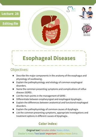
Barrett's Esophagus

A 57-year-old man presents with symptoms of Gastroesophageal Reflux Disease (GORD) and is diagnosed with Barrett's esophagus. Biopsy report is crucial to detect dysplastic changes for adenocarcinoma risk assessment. Another patient, a 51-year-old man, had similar symptoms, highlighting the importance of timely diagnosis and management of Barrett's esophagus.

Download Presentation

Please find below an Image/Link to download the presentation.
The content on the website is provided AS IS for your information and personal use only. It may not be sold, licensed, or shared on other websites without obtaining consent from the author.If you encounter any issues during the download, it is possible that the publisher has removed the file from their server.
You are allowed to download the files provided on this website for personal or commercial use, subject to the condition that they are used lawfully. All files are the property of their respective owners.
The content on the website is provided AS IS for your information and personal use only. It may not be sold, licensed, or shared on other websites without obtaining consent from the author.
E N D
Presentation Transcript
A 57-year-old presents with a history of a retrosternal burning sensation, particularly after large meals, and often on retiring to bed at night. Treatment with antacids has had little effect and he has been referred for endoscopy.
Upper gastrointestinal tract endoscopy reveals reddening of the lower esophageal mucosa There is no evidence of a hiatus hernia. The proximal border of the reddened area is irregular, and this area is biopsied.
1. What is the likely cause of the symptoms? 1. The symptoms of heartburn are suggestive of gastroesophageal reflux disease (GORD), with or without the presence of a hiatus hernia. Other important causes of retrosternal pain should not be overlooked, including cardiovascular causes, especially myocardial ischaemia, as well as other rarer causes including pneumothorax and musculoskeletal pain.
2. What is the final diagnosis? 2. The endoscopic and biopsy appearances confirm a Barrett s oesophagus. This is a metaplastic process which develops as a result of persistent reflux of gastric contents into the esophagus, the normal squamous mucosa being replaced by glandular mucosa of intestinal type.
3. What further information do you require from the biopsy report? 3. It is important to look for dysplastic change in the biopsy which may herald the development of adenocarcinoma.
The patient is a 51-year-old white man who presented 10 years before surgery with a history of heartburn, regurgitation, and epigastric pain. Endoscopy was performed, and a large erythematous area involving the distal esophagus was noted. Biopsy specimens were taken
The patient was treated with antireflux drugs and given a follow-up appointment in 1 year. The patient returned 3 years later, complaining of dysphagia, heartburn, and epigastric pain. Endoscopy was performed again and revealed that the normal white squamous mucosa lining the distal esophagus was replaced by pink mucosa. Biopsy specimens were taken.
Because of the diagnosis of Barrett esophagus, the patient was enrolled in a surveillance program, and yearly endoscopic procedures were recommended. Endoscopy showed extensive Barrett esophagus 6 years before surgery, and biopsy specimens showed features of dysplasia.
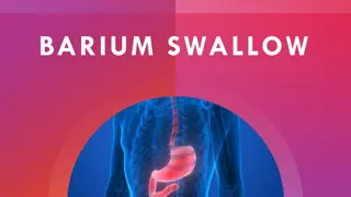
He failed to return for subsequent surveillance endoscopy. The patient was admitted with increasing dysphagia 1 month before surgery. An upper GI series (radiographs) revealed distal narrowing of the esophagus. Endoscopic examination of the esophagus revealed an ulcerating mass in the distal esophagus. A biopsy specimen was obtained.
The patient was taken to surgery, where an esophagogastrectomy was performed.
A 52-year-old male presents with epigastric pain that improves with meals. Endoscopy demonstrates a 2 Cm ulcerated area located 3 cm distal to the pyloric junction. Which of the following is most likely to have made the strongest contribution to the development of this disease? A. Aspirin use B. Chronic antacid use C. Drinking alcohol D. Helicobacter pylori infection E. Smoking
The correct answer is D. The patient has a duodenal peptic ulcer. The strongest risk factor for duodenal peptic ulcer is Helicobacter pylori infection, which is found in almost 100% of these cases (contrast to 70% Infection rate in gastric peptic ulcer). Aspirin use (choice A) and ethanol use (choice C) are more strongly implicated in gastric ulcer disease than duodenal ulcer disease. Chronic antacid use (choice B) is seen as a result of peptic ulcer disease, not as a cause of it. Smoking (choice E) may also be a lesser contributing factor to the development of peptic ulcer.
All of the following are causes of acute peptic ulcer except 1. Severe burns 2. Helicobacter pylori infection 3. Major trauma 4. Zollinger-Ellison syndrome
All of the following are Defensive Factors against gastric ulcer development except A. Mucus B. Bicarbonate C. Bile salts D. Prostaglandins E. Phospholipid
A B 1) H. pylori 2) Phospholipid 3) Drugs (NSAIDs) 4) Mucus 5) bicarbonate 6) Blood flow 7) Acid 8) pepsin 9) Bile salts 10) cell renewal 11) Prostaglandins A. Aggressive Factors B. Defensive Factors
A 49-year-old secretary presents to medical outpatients with a 7- month history of epigastric pain. She has been treated with antacids by her GP, but this has not controlled the symptoms. In the clinic, she complains of epigastric pains which are sharp and burning and radiate her subcostal margin to the right. The pain is worse at night and is relieved by food. On examination, there is epigastric tenderness and clinical signs of anaemia. 1. What is the possible cause of this clinical presentation? Duodenal Peptic ulceration
2. What are the predisposing causes? 1. H PYLORI INFECTION 2. ACID HYPERSECRETION 3. What are the major complications? 3. The major complication is perforation of a vessel with subsequent gastro- intestinal haemorrhage. This can present as either hematemesis, melena or iron deficiency anemia. Other complications include fibrosis and adhesions.
4. What investigations should be performed? Endoscopy 6. What is the treatment? 1. Proton pump inhibitors. 2. H2 receptor antagonists. 3. H. pylori eradication therapy
The patient is a 72-year-old white man with a history of homelessness, chronic obstructive pulmonary disease, chronic alcohol abuse, chronic dementia, and multiple episodes of upper GI bleeding. He was admitted to the hospital with complaints of dizziness, syncope, and abdominal pain. The abdominal examination revealed mild epigastric pain on palpation. The rectal examinations black stool, TEST Hemoglobin Hematocrit Albumin WBC RESULT 4.1 gm/dL 12.9% 2.6 gm/dL 11,900/mm3 <10 mg/dL (normal: <10mg/dL) Ethanol
The patient was admitted to the medical intensive care unit, where a nasogastric tube lavage produced coffee-ground gastric contents that tested positive for blood. He was transfused with 6 U of packed RBCs, which increased his hematocrit to 38% (normal 40% to 52%).
An upper GI tract endoscopy was performed, which showed a large (5 _ 5 cm) gastric ulcer in the antrum along the lesser curvature. Biopsy specimens were taken of the ulcers and surrounding mucosa. Helicobacter pylori