Biochemical Mechanisms of ATP Generation in Respiration
Biochemical mechanisms of ATP generation in higher organisms and bacteria through respiration, fermentation, and photosynthesis. Focus on aerobic and anaerobic respiration, electron transport chain, and oxidative phosphorylation in mitochondria.

Uploaded on Feb 16, 2025 | 0 Views
Download Presentation

Please find below an Image/Link to download the presentation.
The content on the website is provided AS IS for your information and personal use only. It may not be sold, licensed, or shared on other websites without obtaining consent from the author.If you encounter any issues during the download, it is possible that the publisher has removed the file from their server.
You are allowed to download the files provided on this website for personal or commercial use, subject to the condition that they are used lawfully. All files are the property of their respective owners.
The content on the website is provided AS IS for your information and personal use only. It may not be sold, licensed, or shared on other websites without obtaining consent from the author.
E N D
Presentation Transcript
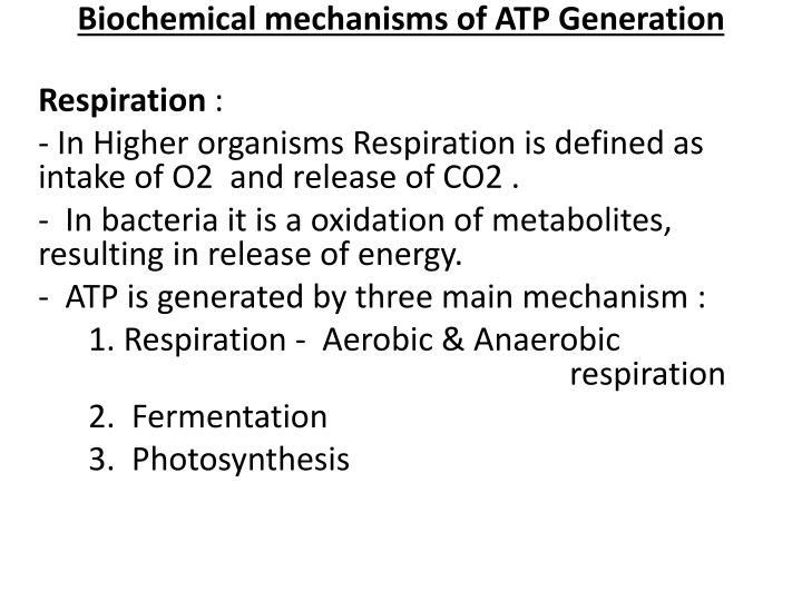
Biochemical mechanisms of ATP Generation Respiration : - In Higher organisms Respiration is defined as intake of O2 and release of CO2 . - In bacteria it is a oxidation of metabolites, resulting in release of energy. - ATP is generated by three main mechanism : 1. Respiration - Aerobic & Anaerobic respiration 2. Fermentation 3. Photosynthesis
Donor Acceptor Donor Aerobic Respiration Organic O2 Inorganic Anaerobic Respiration Organic Inorganic Inorganic Fermentation Organic organic - In Respiration primary donor is oxidizable substrate and terminal acceptor is an inorganic compound either O2 (aerobic) or No3, SO4 or CO3 (anaerobic).
In eukaryotic cells Electron Transport Chain (ETC) & Oxidative Phosphorylation occurs in Mitochondria. - The role of mitochondria in O2 uptake or respiration was demonstrated by Otto Warburg in 1913. - In 1948 by E. Kennedy and Albert Lehninger showed that mitochondria are the site of oxidative phosphorylation in eukaryotes. - The inner membrane bears the components of the respiratory chain and the ATP synthase. - In Bacteria, the plasma membrane plays the same role as inner membrane of mitochondria.
Electron Transport Chain (ETC) ETC of Mitochondria contains large number of electron carrying proteins that acts in sequence to transfer the electrons from reduced substrates to O2. Electron carrying proteins includes : 1. Nicotinamide nucleotides(NAD or NADP ) 2. Flavin nucleotides (FMN or FAD) 3. Ubiquinone or Coenzyme-Q 4. Cytochromes 5. Iron-sulfur proteins
Electron Transport Chain Ketoglurate Malate Pyruvate NAD FP Fe-s CO-Q Cyt-b Cyt-c1 Isocitrate Glutamate FP Succinate Fatty acyl -CoA Cyt-c Cyt-aa3 O2
1. Nicotinamide nucleotides(NAD or NADP) : - Most of the electron pairs enters ETC arise from the action of dehydrogenases utilizes NAD or NADP as acceptors. - Nicotinamide nucleotides linked dehydrogenases catalyze reversible reaction as follows: Reduced substrate + NAD + NADH + H Reduced substrate + NADP + NADPH + H - Most dehydrogenases are specific for NAD as acceptors. - Some are in the cytosol, others are in mitochondria, and still others have mitochondrial and cytosolic isozymes. Oxidized substrate Oxidized substrate
- NAD-linked dehydrogenases remove two hydrogen atoms from their substrates. - One of these is transferred as a hydride ion (:H) to NAD; the other is released as H in the medium. - NADH and NADPH are water-soluble electron carriers that associate reversibly with dehydrogenases. - NADH carries electrons from catabolic reactions. - NADPH generally supplies electrons to anabolic reactions.
2. Flavoproteins : - Flavoproteins are the enzyme with a yellow colored prosthetic group derived from vitamin Riboflavin. - The prosthetic groups are FMN or FAD. Both possess same active site capable of undergoing reversible Oxidation & Reduction. - The oxidized flavin nucleotide can accept either one electron (yielding the semiquinone form) or two (yielding FADH2 or FMNH2).
- Flavoproteins can participate in either one- or two-electron transfers . They serve as Intermediate between two reactions in which two are donated and in which one is accepted. 3. Ubiquinone or Coenzyme-Q : Ubiquinone (coenzyme Q) is a lipid-soluble benzoquinone with a long isoprenoid side chain. - closely related compounds plastoquinone (of plant chloroplasts) and menaquinone (of bacteria).
- Ubiquinone can accept one electron to become the semiquinone radical (QH) or two electrons to form ubiquinol (QH2). - it can act at the junction between a two- electron donor and a one-electron acceptor. - Because ubiquinone is both small and hydrophobic, it is freely diffusible within the lipid bilayer of the inner mitochondrial membrane. - Carries both electrons and protons, it plays a central role in coupling electron flow to proton movement.
4. Cytochrome : The cytochromes are proteins with characteristic strong absorption of visible light, due to their iron containing heme prosthetic groups . - Mitochondria contain three classes of cytochromes, designated a, b, c - distinguished by differences in their light- absorption spectra. - Each type of cytochrome in its reduced (Fe 2 ) state has three absorption bands in the visible range.
- near 600 nm in type a cytochromes, near 560 nm in type b, and near 550 nm in type c. - Cytochromes were first discovered & called histohematins in 1866 by C.MacMunn. - All cytochromes undergo reversible Fe 2 to Fe 3 valance change - The heme cofactors of a and b cytochromes are tightly, but not covalently, bound to their associated proteins; while the hemes of c- type cytochromes are covalently attached through Cys residues.
5. Iron-sulfur proteins : Iron-sulfur proteins ,first discovered by Helmut Beinert. - The iron is present in association with inorganic sulfur atoms or with the sulfur atoms of Cys residues in the protein, or both. - These iron-sulfur (Fe-S) centers range from simple structures with a single Fe atom coordinated to four Cys -SH groups to more complex Fe-S centers with two or four Fe atoms .
Rieske iron-sulfur proteins (named after their discoverer,John S. Rieske) are a variation in which one Fe atom is coordinated to two His residues rather than two Cys residues. - All iron-sulfur proteins participate in one- electron transfers in which one iron atom of the iron-sulfur cluster is oxidized or reduced. - At least eight Fe-S proteins function in mitochondrial electron transfer.
The overall reaction catalyzed by the mitochondrial respiratory chain,where electrons move from NADH, succinate,or some other primary electron donor through flavoproteins, ubiquinone, iron-sulfur proteins, and cytochromes,and finally to O2.
Electron Carriers Complexes : - The electron carriers of the respiratory chain are organized Into four unique complexes. - Complexes I and II catalyze electron transfer to ubiquinone from two different electron donors: NADH (Complex I) and succinate (Complex II). - Complex III carries electrons from reduced ubiquinone to cytochrome- c. - Complex IV completes the sequence by transferring electrons from cytochrome -c to O2.
Complex I: NADH to Ubiquinone : Complex I, also called NADH:ubiquinone oxidoreductase or NADH dehydrogenase, is a large enzyme composed of 42 different polypeptide chains, including an FMN-containing flavoprotein and at least six iron sulfur centers. - Complex I catalyzes two simultaneous and obligately coupled processes: (1) the exergonic transfer to ubiquinone of a hydride ion from NADH and a proton from the matrix, NADH + H + Q NAD + QH2
(2) the endergonic transfer of four protons from the matrix to the intermembrane space. - Complex I is therefore a proton pump driven by the energy of electron transfer, and the reaction it catalyzes is vectorial. - Amytal (a barbiturate drug), Rotenone (a plant product commonly used as an insecticide), and Piericidin A (an antibiotic) inhibit electron flow from the Fe-S centers of Complex I to ubiquinone and therefore block the overall process of oxidative phosphorylation.
- Ubiquinol (QH2, the fully reduced form) diffuses in the inner mitochondrial membrane from Complex I to Complex III, where it is oxidized to Q in a process that also involves the outward movement of H .
Complex - II: Succinate to Ubiquinone : Complex - II is smaller and simpler than Complex I, it contains five prosthetic groups of two types and four different protein subunits. - Subunits C and D are integral membrane proteins. They contain a heme group, heme b, and a binding site for ubiquinone. - Subunits A and B extend into the matrix . They contain three 2Fe-2S centers, bound FAD, and a binding site for the substrate succinate. - The path of electron transfer from thesuccinate- binding site to FAD, then through the Fe-S centers to the Q-binding site.
Complex III : Ubiquinone to Cytochrome c Complex - III, also called cytochrome bc1 complex or ubiquinone:cytochrome c oxidoreductase, couples the transfer of electrons from ubiquinol (QH2) to cytochrome- c with the vectorial transport of protons from the matrix to the intermembrane space. The net equation of Q cycle is : QH2 + 2 cyt c1 (oxidized) + 2H N Q + 2 cyt c1(reduced)+ 4H P
Complex IV : Cytochrome - C to O2 Complex - IV, also called cytochrome oxidase, carries electrons from cytochrome - c to molecular oxygen, reducing it to H2O. - Complex - IV is a large enzyme (13 subunits; Mr 204,000) of the inner mitochondrial membrane. - Bacteria contain a form that is much simpler, with only three or four subunits, but still capable of catalyzing both electron transfer and proton pumping.
- Mitochondrial subunit II contains two Cu ions complexed with - SH groups of two Cys residues in a binuclear center that resembles the 2Fe-2S centers of iron-sulfur proteins. - Subunit -I contains two heme groups, designated a and a3, and another copper ion (CuB). Heme a3 and CuB form a second binuclear center that accepts electrons from heme a and transfers them to O2 bound to heme a3. - Electron transfer through Complex IV is : Cytochrome c CuA center heme - a heme a3 CuB center O2 .
- For every four electrons passing through this complex, the enzyme consumes four substrate H from the matrix (N side) in converting O2 to 2H2O. It also pump one proton outward into the intermembrane space (P side) for each electron that passes through. The overall reaction catalyzed by Complex IV is 4 Cyt c (reduced) + 8H N + O2 4 cyt c (oxidized) + 4H P + 2H2O
Much of this energy is used to pump protons out of the matrix. - For each pair of electrons transferred to O2 - 4 protons are pumped out by Complex-I, 4 by Complex-III and 2 by Complex IV. The equation for the process is : NADH + 11H N + O2 NAD + 10H P + H2O The electrochemical energy due to the difference in proton concentration and separation of charge conserved much of the energy of electron transfer. The energy stored in such a gradient, termed the proton-motive force.
proton-motive force, has two components: (1) the chemical potential energy due to the difference in concentration of a chemical species (H ) in the two regions separated by the membrane. (2) The electrical potential energy that results from the separation of charge when a proton moves across the membrane. - The electrochemical energy in the proton gradient drives the synthesis of ATP from ADP and Pi.
Oxidative phosphorylation : The chemiosmotic model proposed by Peter Mitchell. - According to the model the electrochemical energy inherent in the difference in proton concentration and separation of charge across the inner mitochondrial membrane the proton-motive force drives the synthesis of ATP as protons flow passively back into the matrix through a proton pore associated with ATP synthase.
- Mitchell used chemiosmotic to describe enzymatic reactions that involve, simultaneously, a chemical reaction and a transport process. electrons from NADH and other oxidizable substrates pass through a chain of carriers arranged asymmetrically in the inner membrane. - Electron flow is accompanied by proton transfer across the membrane, producing both a chemical gradient (pH) and an electrical gradient.
- The inner mitochondrial membrane is impermeable to protons; - protons can reenter the matrix only through proton-specific channels (Fo). - The proton-motive force that drives protons back into the matrix provides the energy for ATP synthesis, catalyzed by the F1 complex associated with Fo.
ATP Synthase : - This large enzyme complex of the inner mitochondrial membrane catalyzes the formation of ATP from ADP and Pi. - Also called Complex V, has two distinct components: F1, a peripheral membrane protein, and Fo (o denoting oligomycin- sensitive), which is integral to the membrane. - F1, was identified and purified by Efraim Racker and his colleagues in the early 1960s.
- Isolated F1 catalyzes ATP hydrolysis (the reversal of synthesis) and was therefore originally called F1ATPase. - F1 is unstable at 0 c, relatively stable at R.T. - F1 is a knob like portion, flattened sphere, 8 nm high and 10 nm across, consisting of alternating and subunits arranged like the sections of an orange. (Fig.) - F1 has nine subunits of five different types, with the composition 3 3 . Mol. Wt. 3,80,000
- Each of the three subunits has one catalytic site for ATP synthesis. - the amino acid sequences of the three subunits are identical, their conformations differ. - subunit conformations are designated - ATP, -ADP, and -empty. - subunit making up a central shaft that passes through F1.
- The Fo complex - a proton pore is composed of three subunits, a, b, and c, in the proportion ab2c10 12. Subunit c is a small (Mr 8,000), hydrophobic polypeptide, consisting almost entirely of two transmembrane helices, with a small loop extending from the matrix of the membrane.
Rotational Catalysis : - Paul Boyer proposed a Rotational catalysis mechanism in which the three active sites of F1 take turns catalyzing ATP synthesis. - A given subunit starts in the -ADP conformation, which binds ADP and Pi. - The subunit now changes conformation, assuming the - ATP form. - Finally, the subunit changes to the -empty conformation, which has very low affinity for ATP, and the newly synthesized ATP leaves the enzyme surface.
Another round of catalysis begins when this subunit again assumes the -ADP form and binds ADP and Pi. The conformational changes central to this mechanism are driven by the passage of protons through the Fo portion of ATP synthase. With each rotation of 120, comes into contact with a different subunit, and the contact forces that subunit into the - empty conformation.
The three subunits interact in such a way that when one assumes the -empty conformation, its neighbour to one side must assume the -ADP form, and the other neighbour the -ATP form. One complete rotation of the subunit causes each subunit to cycle through all three of its possible conformations, and for each rotation, three ATP are synthesized and released.
P/O ratio : - Number of inorganic phosphate molecules taken up to phosphorylate ADP per atom of O2 consumed , and is indicative of the efficiency by which coupling takes place. - Most experiments have yielded P/O ratios of between 2 and 3 when NADH was the electron donor, and between 1 and 2 when succinate was the donor. - Number of protons pumped out per pair of electrons are 10 for NADH and 6 for succinate.
- The most widely accepted experimental value for number of protons required to drive the synthesis of an ATP molecule is 4, of which 1 is used in transporting Pi, ATP, and ADP across the mitochondrial membrane. - If 10 protons are pumped out per NADH and if 4 protons required to produce 1 ATP, the proton-based P/O ratio is 2.5 for NADH as the electron donor and 1.5 (6/4) for succinate.


 &")
")

 :")


 the endergonic transfer of four protons from")
")
")
")