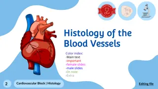
Blood Vessels
Vascular diseases are a leading cause of morbidity and mortality globally, primarily caused by narrowing or weakening of blood vessels. Atherosclerosis, characterized by atheromatous plaques, is a major contributor to cardiovascular events. Learn about risk factors, epidemiology, and pathogenesis of these diseases.

Download Presentation

Please find below an Image/Link to download the presentation.
The content on the website is provided AS IS for your information and personal use only. It may not be sold, licensed, or shared on other websites without obtaining consent from the author.If you encounter any issues during the download, it is possible that the publisher has removed the file from their server.
You are allowed to download the files provided on this website for personal or commercial use, subject to the condition that they are used lawfully. All files are the property of their respective owners.
The content on the website is provided AS IS for your information and personal use only. It may not be sold, licensed, or shared on other websites without obtaining consent from the author.
E N D
Presentation Transcript
Blood Vessels Vascular diseases are responsible for more morbidity and mortality than any other group of human diseases. Vascular pathology result in disease via 2 principle mechanisms:
1- Narrowing or complete obstruction---Progressive or sudden 2- Weakening----Dilatation or rupture.
Normal Vessels - Intima, media, adventitia - Internal elastic lamina - Fenestration - Vasavasorum - External elastic lamina
Atherosclerosis (ATH) Atherosclerosis is characterized by Intimal lesion called atheromas (atheromatousor atherosclerotic plaques), that protrude into the vascular lumina. An atheromatous plaque consists of a raised lesion with a soft, yellow, grumouscore of lipid (mainly cholesterol) covered by a firm white fibrous cap. Besides obstructing the blood flow, atherosclerotic plaque weaken the underlying media.
Atheroma, atheromatousor fibrofatty plaque. Epidemiologic data on ATH are expressed largely in terms of the incidence of the number of deaths caused by ischemic heart disease( IHD).
Atherosclerosis ATH Symptomatic atherosclerotic disease most often involves the arteries supplying heart, brain, kidneys, and lower extremities. Myocardial infarction (Heart attack), cerebral infarction ( stroke) , aortic aneurysm, and peripheral vascular disease ( gangrene of the legs) are the major consequences of ATH.
Epidemiology Atherosclerosis is common in developed countries than Central and South America, Africa and Asia.
Risk Factors Constitutional Risk Factors Age Gender Genetic factors.
Modifiable risk factors Hyperlipidemia. Hypertension Cigarette smoking Diabetes mellitus.
Pathogenesis The response to injury hypothesis: This hypothesis views the atherosclerosis as chronic inflammatory response of the arterial wall to endothelial injury.
Pathogenesis 1- Chronic endothelial injury with resultant endothelial dysfunction, increased permeability, leukocyte adhesion and thrombosis.
Pathogenesis 2-Accumilation of lipoprotein mainly LDL in the vessel wall.
Pathogenesis 3-Monocyte adhesion to the endothelium followed by migration into the intima and transformation into macrophages and foam cells.
Pathogenesis 4-Platelet adhesion
Pathogenesis 5-Factors released from activated platelets, macrophages and endothelium induces SMCs recruitment from media.
Pathogenesis 6- SMCs proliferation and ECM production.
Pathogenesis 7- Lipid accumulation both extracellularly and within the cells.
Morphology 1- fatty streak: Are composed of lipid-filled foam cells but are not significantly raised and so does not affect the blood flow. They begin as multiple minute yellow flat spots which then coalesce into elongated streaks 1cm long and start to appear in the aorta of infants younger than 1 year.
Morphology 2-Atherosclerotic plaques: Are raised masses protrude into the lumen and vary in size from 0.3 to 1.5cm, appear yellow white or red if thrombosis is superimposed. The most common sites are the lower abdominal aorta, coronary arteries, popliteal arteries, internal carotid arteries and vessels of circle of Willis.
Morphology Atherosclerotic Plaque 1- Cells including: SMCs, macrophages, and T cells. 2- ECM: including collagen, elastic fibers and proteoglycans. 3- Intracellular and extracellular lipid. The superficial layer is called fibrous cap and is composed of SMCs and dense collagen. Deep to it is soft necrotic area composed of lipid, debris and foam cells.
Complications 1- Rupture, ulceration or erosion. 2-Hemorrhage. 3- Atheroembolism. 4- Aneurysmal formation


")