Bone Pathology: Central Giant Cell Granuloma and Cherubism Overview
Download Presenatation

Explore the clinical features, radiographic views, histopathology, and treatment options for Central Giant Cell Granuloma and Cherubism in bone pathology. Learn about the characteristics, manifestations, and management of these conditions affecting the mandible and maxilla.

Download Presentation
Please find below an Image/Link to download the presentation.
The content on the website is provided AS IS for your information and personal use only. It may not be sold, licensed, or shared on other websites without obtaining consent from the author. If you encounter any issues during the download, it is possible that the publisher has removed the file from their server.
You are allowed to download the files provided on this website for personal or commercial use, subject to the condition that they are used lawfully. All files are the property of their respective owners.
The content on the website is provided AS IS for your information and personal use only. It may not be sold, licensed, or shared on other websites without obtaining consent from the author.
E N D
Presentation Transcript
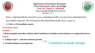
BONE PATHOLOGY CHAPTER 14
Clinical feature Female BONE PATHOLOGY Mandible, anterior portion Asymptomatic nonaggressive aggressive: pain, perforation root resorption, rapid growth CHAPTER 14
Radiographic view Well delineated radiolucent defects Multifocal: hyperparathyroidism cherubism
Histopathology Ovoid to spindle mesenchymal cells Multinucleated giant cells (osteoclast)
Treatment Curretage 11-50% rec Aggressive types
CHERUBISM Chromosome 4
Clinical feature Chrublike faces Eyes upturned to heaven Mandible: painless bilateral exp. posterior areas Maxilla : milder tuberosity
Radiographic view Multilocular, expansile radiolucencies
Histopathology Like CGCG Giant cells : small & focal Cufflike eosinophilic deposites
Treatment & Prognosis Varying degrees of remission after puberty BONE PATHOLOGY CHAPTER 14 Treat or observe Irradiation is contraindicated
Simple Bone Cyst Definition Etiology: trauma-hemorrhage theory Pseudocyst
Clinical feature Common in jaws : mandible molar & premolar area Age: 10-20 asymptomatic
Radiographic view Well delineated radiolucency Domelike projection Vital teeth without resorption
Histopathology Psedocyst----> no ep. Lining Vascular fibrous connective tissue Trabeculae of reactive bone
Diagnosis & Treatment Surgical exploration After 6 months BONE PATHOLOGY CHAPTER 14
Aneurysmal Bone Cyst Intraosseous blood-filled spaces surrounded by connective tissue
Clinical feature unommon in jaws : posterior mandible Age: children & young adults Expansion( rapid ),pain
Radiographic view Unilocular or multilocular radiolucency + cortical exp. & thinning
Histopathology Spaces filled with blood surrounded by cellular fibriblastic tissue no endothelium Giant cells Osteoid & woven bone
Treatment & prognosis Curettage or enucleation cure After 6 months BONE PATHOLOGY CHAPTER 14 Irradiation is contraindicated
Fibro-osseous lesions Definition Developmental Dysplastic Neoplastic
Fibrous dysplasia GNAS1 mutation Time of mutation Monostatic Polyostatic
Monostatic 85% Common in jaws Second decade Painless expansion maxilla
Radiographic view Ground glass Mandible: expansion in buccal,lingual inferior border
Polyostatic FD + caf au lait -----> jaffe-lichtenstein cure +caffe au lait + endocrinopathy ---->Mccune BONE PATHOLOGY CHAPTER 14 albright Mazabraud syndrom
Histopathology Woven osseous trabeculae in cellular fibrous stroma Chinese descript Monotonous pattern
Treatment & prognosis Small lesions cure BONE PATHOLOGY CHAPTER 14 Cosmetic & functional deformity ---> surgery Transformation to osteosarcoma: rare