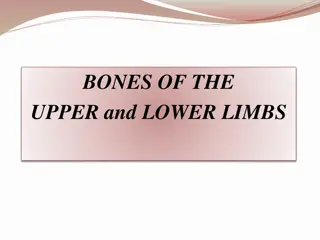
Bones of the Pelvic Girdle and Lower Extremity
This collection of images showcases the bones of the pelvic girdle, including the sacrum, ilium, ischium, pubis, and femur. Detailed views of key structures like the acetabulum, obturator foramen, and greater trochanter are provided. Additional images feature the patella, tibia, fibula, and foot bones, giving a comprehensive visual overview of the bones in the lower extremity.

Download Presentation

Please find below an Image/Link to download the presentation.
The content on the website is provided AS IS for your information and personal use only. It may not be sold, licensed, or shared on other websites without obtaining consent from the author.If you encounter any issues during the download, it is possible that the publisher has removed the file from their server.
You are allowed to download the files provided on this website for personal or commercial use, subject to the condition that they are used lawfully. All files are the property of their respective owners.
The content on the website is provided AS IS for your information and personal use only. It may not be sold, licensed, or shared on other websites without obtaining consent from the author.
E N D
Presentation Transcript
Bones of the Pelvic Girdle And Lower Extremity
Sacrum Ilium Iliac Fossa Pelvic Brim Acetabulum Obturator Foraman Ischium Pubis Pubic Symphasis Pelvis
Coxal Bone Iliac Crest Posterior Superior Iliac Spine Anterior Superior Iliac Spine Anterior Inferior Iliac Spine Posterior Inferior Iliac Spine Acetabulum Greater Sciatic Notch Obturator ForamenIschial Tuberosity Ischial Spine
Greater Trochanter Lateral Epicondyle Inter- trochnteric Line Medial Epicondyle Lesser Trochanter Head Adductor Tubercle Neck Femur - Anterior
Femur - Posterior Lesser Trochanter Gluteal Tuberosity Linea Aspera Medial Epicondyle Medial Condyle Head Lateral Condyle Greater Trochanter Inter- trochanteric Crest Lateral Epicondyle
Patella & Tibia Patella Medial Malleolus Anterior Tibial CrestTibial Tuberosity Medial Condyle Intercondylar Eminence Lateral Condyle
Fibula Lateral Malleolus Head
Proximal Phalanx Middle Phalanx 5 4 Distal Phalanx 3 2 1 Proximal Phalanx Tarsals Metatarsals Phalanges Foot
Cuboid Calcaneous Talus 1st 3rd Navicular 2nd Cuneiform Cuneiform Cuneiform Foot - Tarsals