Brain MRI Measures in Cardiovascular Disease Risk Factors Study
Explore the association between traditional cardiovascular disease risk factors, atrial fibrillation, and brain MRI measures in the Multi-Ethnic Study of Atherosclerosis. The research investigates the impact of age, hypertension, smoking, diabetes, and other factors on brain volumes and white matter injury, shedding light on potential links between cardiovascular health and brain health.

Download Presentation

Please find below an Image/Link to download the presentation.
The content on the website is provided AS IS for your information and personal use only. It may not be sold, licensed, or shared on other websites without obtaining consent from the author. If you encounter any issues during the download, it is possible that the publisher has removed the file from their server.
You are allowed to download the files provided on this website for personal or commercial use, subject to the condition that they are used lawfully. All files are the property of their respective owners.
The content on the website is provided AS IS for your information and personal use only. It may not be sold, licensed, or shared on other websites without obtaining consent from the author.
E N D
Presentation Transcript
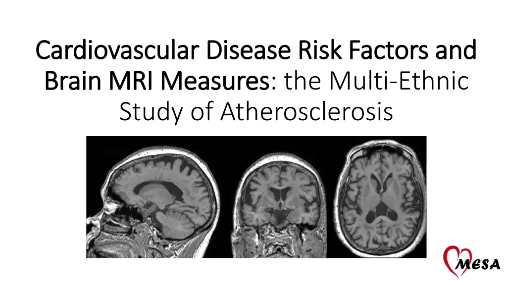
Cardiovascular Disease Risk Factors and Cardiovascular Disease Risk Factors and Brain MRI Measures Brain MRI Measures: the Multi-Ethnic Study of Atherosclerosis
Writing Group Thomas R. Austin R. Nick Bryan Lin Yee Chen Christos Davatzikos Lisa M. Desiderio Guray Erus Philip Greenland Mohamad Habes Barbara N. Harding Timothy M. Hughes W.T. Longstreth Jr. Ilya M Nasrallah Wendy Post Steven J. Shea Susan R. Heckbert
Background Traditional CVD risk factors age, hypertension, smoking, diabetes, physical activity are associated with low gray matter volume and high white matter (WM) lesion volume Most studies include Whites only Clinically-detected atrial fibrillation (AF) is associated with accelerated cognitive decline, but little information about subclinical AF and brain morphologic changes is available
Research Questions 1. Are traditional CV risk factors associated with brain volumes and with three measures of WM injury? 2. Is clinically- and/or monitor-detected AF associated with the same brain MRI measures?
Exposures: Analysis 1 Traditional CV risk factors Age Body Mass Index Smoking Status HDL & LDL Cholesterol Kidney Function - eGFR Systolic BP Hypertension Medication Diabetes
Exposures: Analysis 2 (N = 120) AF: Prevalent atrial fibrillation or flutter Clinically-detected up to E6 (diagnosis codes, Medicare claims, or self-report) OR Zio Patch- detected E6, up to 28-day monitoring Intermittent: prevalent AF but not continuous on Zio Patch (N=100) Continuous: prevalent AF, 100% of time on Zio Patch (N=20)
Analysis Sample N Female (%) Age, yrs, mean(SD) Race/Ethnicity White (%) Chinese-American (%) African-American (%) Hispanic (%) Cigarette Use Never (%) Former (%) Current (%) BMI, mean(SD) HDL Cholesterol, mean(SD) LDL Cholesterol, mean(SD) Systolic BP, mmHg, mean(SD) Hypertension Medication (%) Diabetes (%) Prevalent AF (%) 1,032 53 73 (8) 1,535 with E6 Patch Monitoring 40 15 25 20 Median interval between patch monitoring & brain MRI: 17 months 48 46 6 1,061 with Brain MRI 28 (5) 60 (18) 107 (35) 127 (20) 59 22 12 Analysis Sample 1,032 with complete covariate data
Outcomes: Brain Volumes Volume Measures 1. Total Brain Volume (gray matter + white matter) (mL) 2. Total Gray Matter Volume (mL) 3. Total White Matter Volume (mL)
Outcomes: Brain MRI Measures Measures of White Matter Injury 1. Total WM Lesion Volume (mL) 2. Total WM Fractional Anisotropy (FA) 3. Total WM Apparent Diffusion Coefficient (ADC)
Outcomes: Brain MRI Measures Measures of White Matter Injury 1. Total WM Lesion Volume (mL) 2. Total WM Fractional Anisotropy (FA) 3. Total WM Apparent Diffusion Coefficient (ADC)
Statistical analysis Multivariable linear regression with robust standard errors WM lesion volume log-transformed FA and ADC standardized All models adjusted for MESA site Volume measures adjusted for total intracranial volume (ICV)
Results: CVD Risk Factors & Volumes (Multivariable*) *regression models adjusted for all risk factors above in addition to sex, race/ethnicity, site, and total intracranial volume.
Results: CVD Risk Factors & WM Injury (Multivariable*) * regression models adjusted for all risk factors above in addition to sex, race/ethnicity, site, and total intracranial volume (for WM lesion volume)
Analysis 2 Results: Atrial Fibrillation Regression models adjusted for age, race/ethnicity, sex, site, BMI, smoking, HDL cholesterol, LDL cholesterol, eGFR, SBP, hypertension medication, diabetes status, and total intracranial volume (for volume measures)
Analysis 2 Results: Atrial Fibrillation (cont.) Regression models adjusted for age, sex, race/ethnicity, site, BMI, smoking, HDL cholesterol, LDL cholesterol, eGFR, SBP, hypertension medication, diabetes status, and total intracranial volume (for volume measures)
Limitations Cross-sectional analyses Imperfect AF ascertainment Healthier subset of MESA participants Residual Confounding
Conclusions Age, current smoking, diabetes and hypertension associated with greater WM injury We did not detect an association between kidney function, cholesterol, or BMI and WM injury AF associated with greater WM injury Continuous AF associated with low gray matter volume
Relation to Existing Literature Associations of brain volumes and white matter lesion volume with advanced age, hypertension, smoking, and diabetes are well-established MESA data add to the growing literature on newer measures of WM injury The findings related to AF and WM injury have not been seen in other cohorts.
Next Steps Additional brain MRI measures Region-specific analyses Cerebral microbleeds Cerebral blood flow, vascular reactivity, functional connectivity, infarcts Computer-based pattern classification of brain atrophy: AD & brain aging indices Additional exposures Supraventricular and ventricular ectopy on Zio Patch
Brain MRI Measure Distributions Total White Matter Volume ( l) Total Gray Matter Volume ( l) Total Brain Volume ( l)
Brain MRI Measure Distributions (cont.) Total White Matter Lesion Volume ( l) White Matter Fractional Anisotropy White Matter Apparent Diffusion Coefficient 2.0e-04 1.5e-04 Density 1.0e-04 5.0e-05 0 0 20000 40000 60000 80000 White Matter Lesion Volume - White Matter (Total) Log-Transformed Total White Matter Lesion Volume .3 .2 Density .1 0 2 4 6 8 10 12 wm_wmlvol_log
Correlation Between WM Injury Measures FA & ADC FA & WML Volume ADC & WML Volume .003 .45 .45 Apparent Diffusion Coefficient - White Matter Fractional Anisotropy - White Matter Fractional Anisotropy - White Matter .0028 .4 .4 .0026 .35 .35 .0024 .0022 .3 .3 .0022 .0024 Apparent Diffusion Coefficient - White Matter .0026 .0028 .003 2 4 6 8 10 12 2 4 6 8 10 12 Log-Transformed WML Volume Log-Transformed WML Volume Correlation Coefficient = -0.81 Correlation Coefficient = -0.48 Correlation Coefficient = 0.61
Gender and Race/Ethnicity in Multivariable Model Regression models adjusted for age, race/ethnicity, sex, site, BMI, smoking, HDL cholesterol, LDL cholesterol, eGFR, SBP, hypertension medication, diabetes status, and total intracranial volume (for volume measures)
8-level Categorical Education Included In Adjustment Model *regression models adjusted for all risk factors above in addition to sex, race/ethnicity, site, education, and total intracranial volume.
8-level Categorical Education Included In Adjustment Model (cont.) *regression models adjusted for all risk factors above in addition to sex, race/ethnicity, site, and (for WML Volume) total intracranial volume.
Identification of AF ICD-9/10 No AF AF No AF 936 33 E6 Self- Report AF 30 29 ZioPatch No AF AF No AF 932 30 E6 Self- Report AF 40 19 ICD-9/10 No AF AF No AF 904 47 ZioPatch AF 34 15


")
")

")


")