Breast Disease: Clinical Presentations and Screening Insights
Explore clinical presentations of breast disease, including pain and inflammation, along with insights on mammographic screening for early detection of breast carcinomas. Learn about the likelihood of malignancy and the importance of early diagnosis.

Download Presentation

Please find below an Image/Link to download the presentation.
The content on the website is provided AS IS for your information and personal use only. It may not be sold, licensed, or shared on other websites without obtaining consent from the author. If you encounter any issues during the download, it is possible that the publisher has removed the file from their server.
You are allowed to download the files provided on this website for personal or commercial use, subject to the condition that they are used lawfully. All files are the property of their respective owners.
The content on the website is provided AS IS for your information and personal use only. It may not be sold, licensed, or shared on other websites without obtaining consent from the author.
E N D
Presentation Transcript
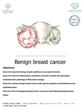
BREAST; STROMAL TUMORS, BENIGN EPITHELIAL LESIONS AND CARCINOMA IN SITU (A) Maram Abdaljaleel, MD Dermatopathologist and Neuropathologist University of Jordan, School of Medicine
Diagnostic pathology book, normal histology, 2nd edition, LINDBERG LAMPS
Diagnostic pathology book, normal histology, 2nd edition, LINDBERG LAMPS
REGARDLESS OF THE SYMPTOM: The underlying cause is benign in >90% of cases. The likelihood of malignancy increases with age: the risk of nipple discharge being due to cancer increases from 7% in women <60 years vs. 30% in women >60. only 10% of palpable masses in women <40 years are carcinomas vs. 60% in women >50.
Of women with cancer: about 45% have symptoms Palpable mass>>>> pain> nipple discharge > inflammatory changes the remainder come to attention through screening tests
MAMMOGRAPHIC SCREENING: detects early, nonpalpable asymptomatic breast carcinomas before metastasis. the average size of invasive carcinomas detected by mammography is about 1 cm, at this stage only 15% will have metastasized to regional lymph nodes The sensitivity and specificity of mammography increase with age due to replacement of the fibrous, radiodense tissue of young women with the fatty, radiolucent tissue of older women
CLINICAL PRESENTATIONS OF BREAST DISEASE: Pain: - cyclic: diffuse, premenstrual edema and swelling. - noncyclic: Localized, ruptured cyst or physical trauma, or infection - Almost all painful masses are benign except for 10% of cases that relates to cancers Inflammation: - causes edematous and erythematous breast. - most often caused by infections (during lactation and breastfeeding). - An important mimic of inflammatory breast cancer
Nipple discharge: - Normal: when small in quantity and bilateral. - Milky discharges (galactorrhea): - are associated with elevated prolactin levels (pituitary adenoma), hypothyroidism, or endocrine anovulatory syndromes, patients taking OCPs, tricyclic antidepressants, methyldopa, or phenothiazines. - Bloody or serous discharges: - commonly due to large duct papillomas and cysts. - During pregnancy, result from the rapid growth and remodeling of the breast. - BUTspontaneous, unilateral, and bloody discharge increases concern for malignancy.
Palpable masses: - 95% are benign - all palpable masses require evaluation. - The most common palpable lesions are cysts, fibroadenomas, and invasive carcinomas - generally detected when they are 2 to 3 cm in size. Gynecomastia: - The only common breast symptom in males. - resulting from an imbalance between estrogens, which stimulate breast tissue, and androgens, which counteract these effects.
INFLAMMATORY PROCESSES: rare caused by infections, autoimmune disease, or foreign body type reactions. Clinically: erythema, edema, pain and focal tenderness. The only infectious agent is Staphylococcus aureus Enters via fissures in nipple skin during the first weeks of breastfeeding lactational abscesses If untreated, tissue necrosis fistula tracks opening onto the skin.
Treatment: antibiotics and continued expression of milk. Rarely, surgical incision and drainage is required. Note: Because inflammatory diseases are rare, the possibility that the symptoms are caused by inflammatory carcinoma should always be considered