Bronchiolitis in Children
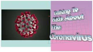
Bronchiolitis is a common lower respiratory tract infection in children under 2 years old, often caused by RSV. It presents with symptoms like cough, wheezing, and fever, and can be managed at home in most cases. However, hospitalization may be necessary in severe cases, especially for infants with risk factors like prematurity or pre-existing conditions. Diagnosis is based on clinical history and examination, with indications for hospitalization including poor feeding, lethargy, and respiratory distress.

Download Presentation

Please find below an Image/Link to download the presentation.
The content on the website is provided AS IS for your information and personal use only. It may not be sold, licensed, or shared on other websites without obtaining consent from the author.If you encounter any issues during the download, it is possible that the publisher has removed the file from their server.
You are allowed to download the files provided on this website for personal or commercial use, subject to the condition that they are used lawfully. All files are the property of their respective owners.
The content on the website is provided AS IS for your information and personal use only. It may not be sold, licensed, or shared on other websites without obtaining consent from the author.
E N D
Presentation Transcript
Bronchiolitis Paula Chilvers GPST2 November 2017
Background Commonest LRTI in children <12m, occurs up to 2yrs old, peak 3-6m Most cases: RSV (Respiratory Syncytial Virus) Oedema of airways widespread narrowing air trapping Majority of cases can be managed at home with support from Primary Care; usually self limiting, lasting 3-7 days Symptoms peak at 4-5 days SAFETY NET Most common cause of hospital admission in infants <6m Can be life threatening, esp if pre-existing cardiac/respiratory disease
Presentation Coryzal prodrome 1-3 days Then, persistent cough Wheeze/crackles on auscultation +/- fever Poor feeding typically after 3 to 5 days Infant <6wks may present only with apnoea
Indications for hospitalisation Poor feeding: <1/2 normal feeds Lethargy Tachypnoea (>70/min) or apnoea Nasal flaring or grunting Moderate to severe chest wall recession SpO2 <93% in air
Risk factors for severe disease Premature birth Age <12w at presentation Pre-existing cardiac/respiratory disease Immunodeficiency
Diagnosis Based on clinical hx and examination Clinical features: - Early symptoms coryzal, non specific - 1-3 days, increasing breathlessness, cough, tachypnoea, varying degrees of respiratory distress - Apnoea esp very young, premature or low birth wt infants - Auscultation: early fine crackles, coarser during recovery; +/- expiratory wheeze - Fever >38.5C in 50% of infants
Diagnosis History - Risk factors eg pre-existing conditions - Feeding pattern duration & completion - Breathlessness/ rapid breathing/ wheeze - Cough, apnoea, cyanotic spells - Wet nappies - Fever? Examination - Degree of distress; circulation, hydration status
Differential diagnosis ?pneumonia if : high fever (over 39 C) and/or focal crackles ?viral-induced wheeze/early-onset asthma older infants and young children with: persistent wheeze no crackles or recurrent episodic wheeze or Personal/family history of atopy Note: Unusual <1 yr old
Investigations Mild cases no Ix required All moderate to severe cases NPA to microbiology for identification of respiratory viruses inc RSV
Investigations Any other Ix are not routine, may be requested after discussion +/- senior review - CXR - FBC - Serum electrolytes if IV therapy required - Blood culture if temp > 38.5C - Capillary blood gas
Treatment NICE: pharmacological agents NOT shown to give benefit above standard supportive care - Oxygen (humidified) single most useful therapy - Airway support CPAP severe respiratory distress/fatigue/apnoea
Treatment Feeding based on degree of tachypnoea, likelihood developing fatigue Bronchodilators should NOT be routinely used (NICE no evidence of definitive benefit). May give short term relief Nebulised 3% hypertonic saline (3HS) questionable benefit
Treatment Antibiotics only if secondary bacterial infection is strongly suspected. Areas of consolidation on CXR not necessarily an indication. Partial R upper lobe collapse quite common in uncomplicated RSV bronchiolitis Corticosteroids no evidence for inhaled or systemic steroids in acute infection Ribavirin no evidence significant benefit RSV prophylaxis Palivizumab for at risk infants
Advice to carer Prevention of cross infection - Incubation 2-8 days, viral shedding 3-8 days (up to 4 wks in young infants) - Handwashing, hygiene - Limit affected individual s contact with others
Safety netting/Advice Trust Guidelines: Review with GP within 7 days of discharge There is strong evidence that smoking increases the risk of admission with bronchiolitis Re-infection may occur - Signs of increased work of breathing - Reduced fluid intake (50-75% of normal/no wet nappy in 12hrs) - Apnoea/cyanosis - Fatigue Red flags Cough resolves in 90% by 3 wks Risk of wheeze increased after bronchiolitis if no FHx atopy, should resolve by age 10 yrs(!)