California Paid Sick Leave Policy Proposal 2025
Effective January 1, 2025, proposed changes to the California Paid Sick Leave Policy for part-time employees include switching to an accrual method, removing waiting periods, allowing carryover of unused hours, and capping usage at 40 hours per calendar year. The impact on students, departments, and the organization is discussed, highlighting the more fair and equitable approach.

Download Presentation

Please find below an Image/Link to download the presentation.
The content on the website is provided AS IS for your information and personal use only. It may not be sold, licensed, or shared on other websites without obtaining consent from the author.If you encounter any issues during the download, it is possible that the publisher has removed the file from their server.
You are allowed to download the files provided on this website for personal or commercial use, subject to the condition that they are used lawfully. All files are the property of their respective owners.
The content on the website is provided AS IS for your information and personal use only. It may not be sold, licensed, or shared on other websites without obtaining consent from the author.
E N D
Presentation Transcript
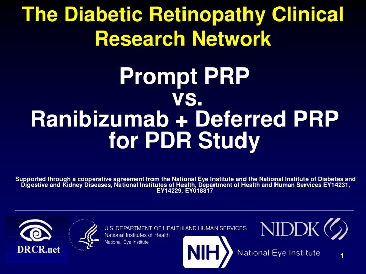
The Diabetic Retinopathy Clinical Research Network Prompt PRP vs. Ranibizumab + Deferred PRP for PDR Study Supported through a cooperative agreement from the National Eye Institute and the National Institute of Diabetes and Digestive and Kidney Diseases, National Institutes of Health, Department of Health and Human Services EY14231, EY14229, EY018817 1
Disclosure Funding/Support: Cooperative Agreement with NEI and NIDDK of NIH, U.S. Department of Health and Human Services. Additional Contributions: Genentech Inc. provided the ranibizumab and clinical site funding. A complete list of all DRCR.net investigator financial disclosures can be found at www.drcr.net. 2
Background Proliferative diabetic retinopathy (PDR): Left untreated, is a leading cause of blindness ~12,000 to 24,000 new cases of diabetic retinopathy-induced blindness each year* Without treatment, ~50% of eyes with high-risk PDR experience severe vision loss within 5 years * CDC: National diabetes fact sheet, 2007 3
Background Panretinal photocoagulation (PRP) has been the treatment for PDR over last 4 decades Substantially reduces risk of severe vision loss, but . . . Inherently destructive Peripheral visual field loss Night vision loss Exacerbation of pre-existing DME Not perfect: 5% severe vision loss (worse than 5/200 at 2 consecutive visits) despite PRP Anti-VEGF, when given for DME, decreases risk of diabetic retinopathy worsening and increases chance of improved retinopathy level 4
Study Design Randomized, multi-center clinical trial (55 Sites) Participants meeting all of the following criteria: Age 18 years with Type 1 or type 2 diabetes Study eye(s) meeting all of the following criteria (a participant can have 2 study eyes): PDR No history of PRP Best corrected visual acuity letter score 24 (~Snellen equivalent 20/320 or better) Eyes with or without central-involved DME were eligible Primary Objective: Compare the efficacy and safety of PRP with that of intravitreous ranibizumab (0.5-mg in 0.05 mL) for proliferative diabetic retinopathy (PDR) 5
Primary Question Is visual acuity using ranibizumab for PDR not worse than treatment with PRP at 2 years? Non-inferiority margin of 5 letters Secondary Question Are there potential benefits of ranibizumab on: Vision throughout follow-up (area under the curve) Peripheral vision Macular edema Incidence of vitrectomy 6
Follow-up Schedule PRP group: Visits every 16 weeks* Ranibizumab group: Visits every 4 weeks to assess for PDR treatment Baseline to 1 Year Both groups simultaneously evaluated for DME treatment PRP group: Visits every 16 weeks* Ranibizumab group: Visits every 4wk to 16wk to assess for PDR treatment 1 Year to 2 Years Interval is extended if injections for PDR and DME deferred ( Defer and Extend ) *Eyes with DME could be seen more frequently for DME treatment as needed. 7
PDR Treatment Schedule: Ranibizumab Group Year 1 6 initial injections q4 weeks One exception: if no neovascularization (NV) at 4-month or 5-month visit, then injection withheld Starting at 6-month visit: Inject if NV improved compared with any previous 3 consecutive visits where injection given Withhold injections if NV stable over previous 3 consecutive injections After injection withheld, resume injections if NV worsens 8
PDR Treatment Schedule: PRP Group Prompt PRP- Initial 1 to 3 sittings within 8 weeks of randomization Standard laser initial full session = 1200 to 1600 burns Automated pattern initial full session = 1800 to 2400 burns Ranibizumab required for eyes with central involved DME and vision loss at baseline. If the size or amount of NV increased following initial PRP, then additional PRP could be given. 9
DME Treatment Schedule: Ranibizumab or PRP Groups Eyes in both groups could receive 0.5-mg ranibizumab for DME treatment At randomization, treatment was required for central DME with visual acuity 20/32 or worse defined as Baseline DME throughout remainder of presentation Otherwise initiation and retreatment with ranibizumab for DME at follow-up was at investigator discretion DRCR.net protocol comparing ranibizumab with prompt or deferred focal/grid laser with focal/grid laser (Protocol I) retreatment regimen provided as guideline Focal/grid laser at investigator discretion 10
Randomization Participants: N = 304 Eyes: N = 394 Ranibizumab Group N = 191 PRP Group N = 203 Baseline 2-Years N = 160 (84%) N = 168 (83%) 2-Years Excluding Death N = 86% N = 88% Median (Quartiles) No. Visits over 2 years N = 16 (9, 22) N = 22 (18, 24) 11
Baseline Characteristics Ranibizumab Group (N = 191) 52 43% PRP Group (N = 203) 51 45% Age (yrs) Median Women Race White Type 2 diabetes Duration of Diabetes (yrs) Median HbA1c (%) 52% 73% 18 8.6 50% 76% 17 8.9 12
Ocular Baseline Characteristics Ranibizumab Group (N = 191) PRP Group (N = 203) Mean visual acuity letter score (~Snellen Equivalent) 75.0 (20/32) 75.2 (20/32) 20/25 or better 20/32 to 20/40 46% 34% 46% 33% 20/50 to 20/100 16% 17% 20/125 to 20/320 5% 4% 13
Ocular Baseline Characteristics Ranibizumab Group (N = 189) 262 66% 19% 15% Required ranibizumab at baseline PRP Group (N = 201) 249 67% 26% 7% Mean OCT CST* ( m) < 250 m 250 to 349 m 350 m Presence of central- involved DME with VA loss** 22% 23% *OCT values are Stratus equivalents **Eyes with visual acuity letter score 78 (20/32 or worse) ANDOCT CST machine and gender specific thresholds
Ocular Baseline Characteristics Ranibizumab Group (N = 189) PRP Group (N = 199) Diabetic Retinopathy Severity by Reading Center NPDR 10% 13% Mild to moderate PDR 52% 49% High risk PDR to advanced PDR 38% 37% There were 46 eyes (12%) for which NV was not identified by the reading center on the submitted color images or quality precluded identification. In 29 of these cases (63%), subsequent review of additional images (e.g. FA) confirmed NV, leaving 17 (4%) of 394 subjects with no photographic documentation of PDR. 15
Treatment For Proliferative Diabetic Retinopathy 16
PRP Group Baseline PRP Overall (N = 203) Completed initial full PRP 98% Performed in 1 Sitting 54% 17
PRP Group Additional PRP Overall (N = 203) Eyes given additional PRP (after completing initial full PRP) 45% Distribution of timing to additional PRP From completion of initial full PRP: median time to additional PRP ~7 months 18
Ranibizumab Group # of Ranibizumab Injections Eyes With Baseline DME (N = 36) Eyes Without Baseline DME (N = 133) Prior to 1-year Visit (Max possible = 13) Median Mean 9 7 8.9 6.9 Prior to 2-year visit (Max possible= 26) Median Mean 14 13.3 10 10.1 Note: 97% of protocol-required injections for PDR were given 19
Ranibizumab Group Received PRP for PDR Overall N = 191 Received PRP* before 2 years 12 (6%) *1 met failure criteria, 1 with Protocol Chair approval, 1 without Chair approval, 8 during vitrectomy (e.g., via endolaser), and 1 by non-study physician 20
Treatment For Diabetic Macular Edema 21
PRP Group Ranibizumab Treatment for DME Timing of first ranibizumab injection for DME Overall (N = 203) 47% Never 35% Baseline 18% Follow-up 22
PRP Group: # of Ranibizumab Injections for DME Eyes with baseline DME (N = 42) Eyes without baseline DME (N = 135) Prior to 1-year Visit (Max = 13) Median 5 0 Mean 5.7 1.4 Prior to 2-year Visit (Max = 26) Median 9 0 Mean 9.1 2.2 23
Additional Treatment for DME Ranibizumab Group PRP Group 10% Focal/grid laser 8% Received 1 or more alternative treatment <1% 2% 24
Efficacy 25
Mean Change in Visual Acuity 15 Mean Visual Acuity Change 10 (Letter Score) 5 0 -5 0 16 32 52 68 84 104 Visit Week 27 Outlying values were truncated to 3 SD from the mean
Mean Change in Visual Acuity 15 Mean Visual Acuity Change 10 (Letter Score) 5 0 + 0.2 -5 0 16 32 52 68 84 104 Visit Week PRP Group N = 168 N = 203 28 Outlying values were truncated to 3 SD from the mean
Mean Change in Visual Acuity 15 2-Year Adjusted Mean Difference: +2.2 letters 95% Confidence Interval: (-0.5, +5.0) (Meets pre-specified non-inferiority criterion: lower bounds of the 95% CI of -0.5 letters was greater than the non-inferiority limit of -5.0 letters) Mean Visual Acuity Change 10 (Letter Score) 5 + 2.8 0 + 0.2 -5 0 16 32 52 68 84 104 N = 191 N = 203 Visit Week N = 160 N = 168 Ranibizumab Group PRP Group 29 Outlying values were truncated to 3 SD from the mean
Mean Change in Visual Acuity Area under the Curve Analysis 15 Adjusted Mean Difference over 2 years (AUC): +4.2 P-value<0.001 95% Confidence Interval: (+3.0, +5.4) Mean Visual Acuity Change 10 (Letter Score) 5 + 4.5 0 -0.3 -5 0 16 32 52 68 84 104 N = 160 Visit Week N = 191 N = 203 N = 168 Ranibizumab Group PRP Group 30 Area under the curve (AUC) analysis: Pre-planned secondary outcome
Mean Change in Visual Acuity Subset that Completed 2 Yrs 15 Mean Change in Visual Acuity 10 (Letter Score) 5 + 2.8 0 + 0.2 -5 0 16 32 52 68 84 104 Visit Week N = 160 of 191 N = 168 of 203 Ranibizumab Group PRP Group 31
Mean Change in Visual Acuity: Eyes Baseline DME 15 Mean Visual Acuity Change 10 (Letter Score) +7.9 5 +1.9 0 2-Year Adjusted Mean Difference: +3.0 letters 95% Confidence Interval: (-4.2, +10.3) -5 0 16 32 52 68 84 104 N = 33 N = 37 Visit Week N = 42 N = 46 Ranibizumab Group PRP Group 32
Mean Change in Visual Acuity Eyes Without Baseline DME 15 2-Year Adjusted Mean Difference: +1.4 letters 95% Confidence Interval: (-1.5, +4.4) Mean Visual Acuity Change 10 (Letter Score) 5 +1.8 0 -0.5 -5 0 16 32 52 68 84 104 N = 126 N = 147 N = 155 Visit Week N = 130 Ranibizumab Group PRP Group 33
Mean Change in Visual Acuity Stratified by Baseline DME Without Baseline DME With Baseline DME 14 Mean Visual Acuity Change 12 10 +7.9 (Letter Score) 8 6 4 +1.8 +2 2 0 - 0.5 -2 -4 0 16 32 Visit Week 52 68 84 104 N = 33 N = 37 0 16 32 Visit Week 52 68 84 104 N = 126 N = 147 N = 155 N = 42 N = 46 N = 130 Ranibizumab Group PRP Group 34 *Outlying values were truncated to 3 SD from the mean
10 Letter Score Improvement* 60 50 Percentage of Eyes (%) 43% 40 36% 30 20 10 2-Year Adjusted Difference: 6% 95% Confidence Interval: (-8%, +21%) 0 0 16 32 52 68 84 104 N = 81 N = 86 N = 104 N = 110 Visit Week Ranibizumab Group PRP Group 35 *Baseline VA 20/32 or worse
10 Letter Score Worsening 60 2-Year Adjusted Difference: -4% 95% Confidence Interval: (-11%, +2%) 50 Percentage of Eyes (%) P= # 40 30 20 14% 10 9% 0 0 16 32 52 68 84 104 N = 160 N = 168 Visit Week N = 191 N = 203 Ranibizumab Group PRP Group 36
15 Letter Score Worsening 60 2-Year Adjusted Difference: -2% 95% Confidence Interval: (-8%, +3%) 50 Percentage of Eyes (%) 40 30 20 10% 10 8% 0 0 16 32 52 68 84 104 N = 160 N = 191 N = 203 Visit Week N = 168 Ranibizumab Group PRP Group 37
Peripheral Visual Field Outcomes 2-Year Visit Humphrey Visual Field 30-2 + 60-4 Ranibizumab Group (N = 58) PRP Group (N = 57) Cumulative Point Score Change from Baseline -23 -422 Mean 372 dB (P<0.001) Difference (P-Value) Mean Deviation Change from Baseline -0.08 -2.50 Mean 2.2 (P< 0.001) Difference (P-Value) 38
Optical Coherence Tomography Central Subfield Thickness 39
Central Subfield Thickness Change: 2-Year Visit Ranibizumab Group PRP Group Mean Change ( m) Adjusted Difference (P-value) -47 -3 -45 m (P < 0.001) Eyes with Baseline DME Mean Change ( m) Adjusted Difference (P-value) -48 -153 -54 m (P= 0.006) Eyes without Baseline DME Mean Change ( m) Adjusted Difference (P-value) +10 -18 -31 m (P < 0.001)
Proportion of Eyes Developing Center Involved DME with Vision Impairment (Eyes without Baseline DME and Vision Impairment) 2-Year Adjusted Difference: 19% 95% Confidence Interval: (10% to 28%) P-value < 0.001 N = 155 N = 147 41
Complications of PDR Ranibizumab Group (N = 191) 6% PRP Group (N = 203) P-value 10% Any retinal detachment 0.08 2% 3% Neovascular glaucoma 0.50 1% 1% Iris neovascularization 0.96 27% 34% Vitreous hemorrhage 0.09 4% 15% Vitrectomy < 0.001 43 PDR = proliferative diabetic retinopathy
Diabetic Retinopathy: 2-Year Visit Ranibizumab Group (N = 142) Prompt PRP Group (N = 148) P- value Fundus Photos Graded by Reading Center* 0.41 35% 30% - No PDR 23% 24% - Regressed NV 42% 46% - Active NV * Only includes eyes with active NV at baseline 44
Diabetic Retinopathy Improvement: Ranibizumab Group (Note: Cannot determine for PRP Group) Ranibizumab Group 2-Year Visit N = 142 Eyes improving 2 or more steps in DR severity on fundus photos 47% 45
Safety 46
Ocular Adverse Events* Ranibizumab Group N = 191 0.5% PRP Group N = 203 P-value 0% -- Endophthalmitis 1% 4% 0.02 Inflammation 0% 0% -- Retinal Tear 2% 9% 6% 13% 0.06 0.16 Cataract Surgery Elevation in IOP 47 *Event defined as occurring at least once through 2 years.
Systemic Adverse Events APTC Events* One Study Eye Ranibizumab Group N = 102 PRP Group N = 114 2- Study Eyes N = 89 2% 3% Non-fatal MI 2% 1% 2% Non-fatal stroke 4% Vascular/ Unknown Death Any APTC event P = 0.80 4% 4% <1% 8% 9% 6% *Anti-platelet Trialists` Collaboration defined events. Occurring at least once through 2 years. 48
Pre-Specified Systemic Adverse Events* One Study Eye 2- Study Eyes N = 89 Ranibizumab Group N = 102 PRP Group N = 114 P- value 4% 6% Death (any cause) 4% 0.70 42% 47% Hospitalization 35% 0.20 Serious adverse event 43% 48% 37% 0.26 16% 25% Hypertension 18% 0.23 49 *Occurring at least once through 2 years
MedDRA Systems (P value < 0.05) One Study Eye N = 26 Systems 2- Study Eyes N = 89 Ranibizumab Group N = 102 PRP Group N = 114 P- value % Per Participant Cardiac 13% 18% 5% 0.01 Endocrine 20% 25% 11% 0.02 Infections/ infestations 28% 27% 14% 0.02 Respiratory 35% 46% 30% 0.04 Skin 20% 24% 11% 0.03 Surgical 8% 24% 16% 0.01 50


")