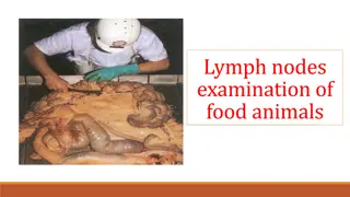
Explore the significance of carcass lymph nodes in meat hygiene, their drainage patterns, and their role in assessing diseases like T.B. Discover how lymph nodes in the carcass are key indicators of the animal's health status.
Please find below an Image/Link to download the presentation.
The content on the website is provided AS IS for your information and personal use only. It may not be sold, licensed, or shared on other websites without obtaining consent from the author. If you encounter any issues during the download, it is possible that the publisher has removed the file from their server.
You are allowed to download the files provided on this website for personal or commercial use, subject to the condition that they are used lawfully. All files are the property of their respective owners.
The content on the website is provided AS IS for your information and personal use only. It may not be sold, licensed, or shared on other websites without obtaining consent from the author.
Definition: they are normally regarded as those lymph nodes which remain on or in the carcass after dressing & include the renal and supramammary L.n. therefore, they comprise all lymph nodes of the body with exception of those of the head and those removed with the thoracic & organs during evisceration. The style of dressing affected whether or not certain lymph nodes will be found on the dressed carcass. Sometimes the dorsal & ventral mediastinal sternal , anterior sternal & prpectoral are removed during evisceration, therefore the 4 L.n. should not be regarded as carcass L.n.
Carcass L.n may drain; 1. Exclusively muscle " meat lymph nodes"; e.g. popliteal, axillary and prescapular L.n. 2. Exclusively organs: e.g. renal L.n. important in the judgment of T.B, because infection is only through hematogenous route. 3. Exclusively skin; precrural L.n.
Meat lymph nodes They have special interest in meat hygiene, they mainly receive lymph from muscles, tendons, skeleton & joints. They are exclusively in bacteriological examination of meat. In cattle, the prescapular, axillary, popliteal& precrural l.n are considered caracss L.n. They are the most important in the judgment of T.B.
Affections of the lymphatic system: The lymph are frequently exhibit various stages of inflammation or degeneration. Consequently, the examination of the lymph nodes of the carcass and organs is the most valuable guide to the nature 7 extent of septicaemic progress in the animal body, and it is also of the greatest value in the judgment of T.B in the carcass.
2. Lymphadenitisor inflammation of the lymph nodes Acute form: Manifested by swelling and oedema of the L.n, which is softer and may be congested, staw berry as in case of swine erysiplas or deeply congested in case of anthrax. Chronic form: Manifested by the development of fibrinous C.T in the affected lymph node which become enlarged and indurated. Such changes are consequently observed in caseous lymphadenitis in sheep, actinobacillosis in cattle & T.B.
2. Parasitic affections: Parasitic affections of lymph nodes are not uncommon. Linguatula larvae or immature fasciola may be found in the mesenteric L.n of the cattle. Also, cysticercus & hydatid cysts may be encountered in the lymph nodes. These parasitic affections tend to undergo caseation and calcification and may be therefore be confused with T.B., Typical greenish color of the caseated parasitic foci together with the case with which they can be shelled out from the affected lymph nodes, are reliable methods of differentiation.
3. Fatty changes: Pathological fatty changes observed in the supramammary L.n of the cow, and seen as white streaks and opaque white spots which are clearly defined against the normal gray translucent lymph node tissue. 4. Lymphadenomatous growth: Characterized by progressive enlargmnet of the lymphoid tissue as in case of lymphatic leukaemia.
5. Pigmentary changes: There are certain pigmentary changes seen particularly in the bronchial L.n, as a result of inhalation of dust and coal particles & this condition is known as anthracosis .
Haemolymph nodes" accessory spleen": These are deep red or almost black in color, oval in shape and up to pea in size, but differ from lymph nodes in their anatomical structure & in the absence of afferent and efferent lymphatic vessels. Haemolymph nodes are supplied by arteries which break up in the gland substance &discharge their blood into tissue space; in this respect these nodes resemble to the spleen, and may be described as accessory spleens .
Haemolymph nodes They contain numerous white blood cells together with red blood corpuscles in various stages of disintgeration, this process being responsible for the red coloration of the nodes. They are larger and more numerous in animals suffering from anaemic conditions.