Cell Division in the Onion Root Tip
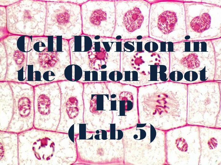
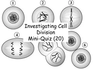
Cell division in the onion root tip is a crucial process for growth and repair. The cell cycle consists of interphase, mitosis, and cytokinesis, each with distinct phases like prophase, metaphase, anaphase, and telophase. These stages involve intricate changes in chromatin, chromosomes, centrioles, and spindle fibers. Mitosis and meiosis play vital roles in cell replication for tissue renewal and gamete production. Understanding these processes is key to comprehending cellular development and function.

Download Presentation

Please find below an Image/Link to download the presentation.
The content on the website is provided AS IS for your information and personal use only. It may not be sold, licensed, or shared on other websites without obtaining consent from the author.If you encounter any issues during the download, it is possible that the publisher has removed the file from their server.
You are allowed to download the files provided on this website for personal or commercial use, subject to the condition that they are used lawfully. All files are the property of their respective owners.
The content on the website is provided AS IS for your information and personal use only. It may not be sold, licensed, or shared on other websites without obtaining consent from the author.
E N D
Presentation Transcript
Cell Division in the Onion Root Tip (Lab 5)
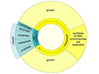
Phases of the Cell Cycle The cell cycle consists of Interphase normal cell activity cell division INTERPHASE S phase Growth (DNA synthesis) G 1 Growth G2
Interphase Interphase : It is a long, metabolically active phase between two successive mitotic cell division. Structural change:- 1- Extended and condensed threads of chromatin material. 2- Intact nucleolus 3- continuous nuclear envelope 4- the cytoplasm of the cell consist of two pairs of centrioles, from which rays of microtubes extend to form a structure called asters, leads to formation of mitotic spindle fibers.
The Cell Division The human cells are replicated by two processes : nuclear division ( mitosis or meiosis ) and cytoplasmic division ( cytokinesis). Mitosis The body cells divide for growth, repair and renew the body tissue. Cytokinesis Meiosis The germ cells divide to produce gametes for reproduction Cytokinesis
The stages of mitosis Prophase It is the longest phase. During this phase the Chromosomes condense and thicken, each duplicated chromosome appear as two identical sister chromatids The centrioles move to the opposite sides of the cell to creat the mitotic spindle fibers Nucleolus and Nuclear membrane disintegrate and disappear
Metaphase During metaphase the following changes take place:- 1- shortening and arrangement of the chromosomes in the middle part of the cell, in an area called equatorial plate( quarter) or metaphase plate. 2- Attachment of microtubules of the spindle fibers to the poles of the cell on one side and to the centromere of the chromosomes on the other side. The point of attachment between the microtubules and the centromere is known as kinetochore.
Anaphase 1- Splitting the centromeres of the chromosomes and separating the chromatin from each other to form the daughter chromosomes, the newly formed chromosomes move to the opposite sides of the cell . 2- shortening of the mitotic spindle fibers. Animal Cell Plant Cell
Telophase 1- Extension of the chromosomes of the daughter cells to become less visible due to decondensation of their molecules to become chromatin material. 2- Nucleolus and nuclear membrane reappears 3- The spindle fibers disappear 4-Finally appearing of a contractile ring in the cleavage groove of the cell to assist in constriction of the plasma membrane during cytokinesis process. Animal Cell Plant Cell
Examining root tip cells Material 1- Onions root tips 2- Microscope 3- Microscope slides and cover slips 4- Toluidine blue or aceto-orcein 5- 1M hydrochloric acid 6- Distilled Water
Procedure 1. Submerge the base of an onion in water at a constant temperature to grown roots for 48 hours. 2. Obtain an onion root and cut off the bottom 1 or 2 mm of the root tip and place it on a Slide. 3. Add a very small drop of 1M HCL acid to the root tip on the slide and let it sit for 4 min. 4. Using a paper to soak the HCL away from the root tip. 5. Cover the root tip with a drop staining solution and let stand for 2 minutes.
Procedure 6. Remove excess stain. Add one drop of water and gently lay a cover slip over the root tip. 7. Using a pencil eraser, carefully apply pressure to the cover slip area to squash and spread the root tip tissue. 8. Use the low power objective on your microscope to look for thin layers of cells and then use the 40X power objective to observe mitotic stages in individual cells. 9. Identify chromosomes at the various stages of mitosis in the water treated root tips. Make sketches of the stages observed.