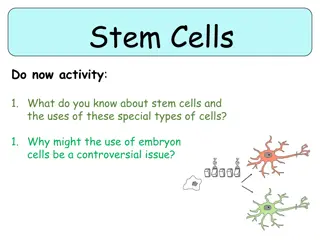
Cells of Acute Inflammation
Acute inflammation is a protective mechanism in response to injurious agents in the body, characterized by vascular changes and specific cell types. Learn about the types, features, and cardinal signs of acute inflammation, as well as the key cells involved, such as Neutrophils.

Download Presentation

Please find below an Image/Link to download the presentation.
The content on the website is provided AS IS for your information and personal use only. It may not be sold, licensed, or shared on other websites without obtaining consent from the author.If you encounter any issues during the download, it is possible that the publisher has removed the file from their server.
You are allowed to download the files provided on this website for personal or commercial use, subject to the condition that they are used lawfully. All files are the property of their respective owners.
The content on the website is provided AS IS for your information and personal use only. It may not be sold, licensed, or shared on other websites without obtaining consent from the author.
E N D
Presentation Transcript
Cells of Acute Inflammation
INTRODUCTION TO INFLAMMATION The reaction of vascularized tissue or circulatory system against injurious agent in living body Reaction can be generalised or localised Mostly protective mechanism Few cases- harmful, Ex- hypersensitivity reaction
Etiological Agent Physical-thermal injury, electric current, x- rays etc. Chemical-acid, toxic gases etc. Biological bacteria, virus, parasite etc. Nutrition Immunological- Ag-Ab complexes Necrotic tissue
TYPES OF INFLAMMATION 2 types 1.Acute Inflammation Due to early response by the body Short duration 2.Chronic Inflammation Occurs after delay Longer duration Characterised by chronic inflammatory cells
Features of Acute & Chronic Inflammation Acute Inflammation Chronic Inflammation Short duration (hr., days) Long duration(week, month, years) Irritant is severe Irritant is of low intensity Marked vascular changes Vascular changes are less prominent Profuse exudation Exudation scacity Soft consistency Firm consistency No or slight proliferation of CT, BV & epithelium More proliferation of CT, BV & epithelium Predominantly neutrophils Predominantly lymphocytes and macrophages
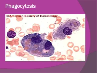
1. NEUTROPHIL Also called as Polymorphs or Polymorphonuclear leukocytes(PMNL) Range:20-30% of DLC Size-12-15 micron Spend <48 hrs in circulation Heterophil-rabbit, guinea pig, domestic fowl First line of cellular defence Neutrophilia -inc. No. Of neutrophils Neutropenia dec. No. Of neutrophils
Microphages of Metchnikoff Fxn : Selective but highly phagocytic Bacterial infection more recruitment of neutrophils Main component of purulent(pus) exudates Granulocyte Colony-Stimulating Factor(G-CSF) stimulate growth of neutrophil
Schilling Index Helps in determining the immature form of neutrophil in blood Shift to left- more immature neutrophils Shift to right- more mature neutrophils
2.Eosinophil Range- 4-6% of DLC Bilobed, Very short lived Parasitic infestation more recruitment Allergic rxn inc. No. Of eosinophil in blood Size-10-20 micron Fxn: Killing of foreign body & allergen Activated eosinophil rich source of leukotrienes- esp. Leukotriene-C4 + PAF
Eosinophil specific granules contain: i.) Major basic protein- toxic to parasite + lysis of epithelial cells ii.) Eosinophilic cationic protein(ECP)- directly toxic to epi. Cells iii.)Eosinophil peroxidase- tissue damage by oxidative stress Have mild phagocytic property Eosinophilia-inc. No. Eosinopenia- dec. No.
3. BASOPHIL/MAST CELL Range: 0.5-1% of DLC Nucleus: round, filled with bluish granules, fragmented nucleus Involved in Type-I hypersensitivity due to histamine secretion from mast cells which have role in allergy Size: 12-15 micron Non phagocytic property Fxn: produces histamine, serotonin & heparin
Basophil contain a no. Of basophilic membrane bound granules Histamine Eosinophil chemotactic factor(ECF) Neutrophil Chemotactic factor(NCF) Granule matrix derieved mediators-heparin Neutral proteases- Tryptase
Property Mast cell Basophil B.M. Mesenchymal cells Produce Blood Around tissues Found in 12-15 micron >30 micron Size Bilobed Without lobes Nucleus Many Few Cytoplasmic granules Mitosis Absent Present