Cellular Elements and Functions in Connective Tissue Biology
Diverse cell types in connective tissue such as fibroblasts, cementoblasts, immune system cells, and more. Learn about the ground substance and its vital functions in maintaining connective tissue health. Discover the unique characteristics and roles of these cellular elements in the periodontal ligament (PDL) and associated structures.

Download Presentation

Please find below an Image/Link to download the presentation.
The content on the website is provided AS IS for your information and personal use only. It may not be sold, licensed, or shared on other websites without obtaining consent from the author.If you encounter any issues during the download, it is possible that the publisher has removed the file from their server.
You are allowed to download the files provided on this website for personal or commercial use, subject to the condition that they are used lawfully. All files are the property of their respective owners.
The content on the website is provided AS IS for your information and personal use only. It may not be sold, licensed, or shared on other websites without obtaining consent from the author.
E N D
Presentation Transcript
Cellular elements 4 Types - connective tissue cells (Fb, Cb, Ob) - epithelial rest cells -immune system cells - cells associated with neurovascular elements
Connective tissue cells Fibroblast- - most common cells in the PDl - ovoid or elongated cells and exhibit pseudopodia like process - synthesize collagen and have the capacity to phagocytose old collagen fib and degrade them by enzyme hydrolysis.
Cementoblast - They are the cells responsible for secreting the organic matrix of cementum in the PDL. Osteoblast - They are found on the surface of the alveolar bone.
Epithelial rest cells - Epithelial rest of Malassez are considered as remnants Hertwigs root sheath which disintegrates during root development. - Most numerous in apical and cervical areas. - when stimulated, may participate in the formation of periapical cyst and lateral root cyst.
Immune system cells Include neutrophils, lymphocytes, macrophages, mast cells and eosinophils. These cells as well as those associated with neurovascular elements, are similar to the cells in other connective tissues.
Ground substance GAG (glycosaminoglycans) - Hyaluronic acid - Proteoglycans GP (glycoproteins) - fibronectin - Laminin Water (70%)
Functions of ground substance Helps nutrients, and metabolites, to and from CT cells, thus is essential for the maintenance of the normal function of CT. in transportation of water, electrolyte,
PDL may also contain calcified masses called cementicles which are adherent to detached from root surface. Cementicles may develop from calcified epithelial rest, around small spicules of cementum or alveolar bone traumatically displaced in to PDL, from calcified sharpey s fib, and from calcified thrombossed vessels in PDL.
Functions of PDL 1. Physical 2. Formative 3. Remodelling 4. Nutritional 5. Sensory
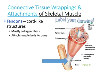
Physical function 1. Provision of soft tissue casing to protect the vessel and nerves from injury to mechanical forces. 2. Transmission of occlusal forces to the bone. 3. Maintenance of gingival tissues in their proper relationship to the teeth. 4. Resistance to the impact of occlusal forces (shock absorption).
SHOCK ABSORPTION 2 theories- -Tensional theory -Viscoelastic system theory
Formative and remodeling function PDL have the regenerative capacity in providing the cell lineage namely ob, cb and fb. Thus it help in the formation and resorption of cementum and bone during physiological tooth movement and repair of injuries.
Nutritional and sensory function PDL supplies nutrients to the cementum, bone and gingiva by way of blood vs and also provide lymphatic drainage. The PDL is abundantly supplied with sensory nerve fib capable of transmitting tactile, pressure and pain sensations by the trigeminal pathway.
There are 4 types of neural terminations are present in PDL 1. Free ending- which have tree like configuration and carry pain sensation 2. Ruffinian corpuscles- Mechanoreceptors (in apical areas) 3. Meisners corpuscles- Mechanoreceptors (in midroot areas) 4. Spindle like receptors- Pressure and vibration receptors/
Homeostasis With the presence of both formative and resorptive activity the PDL provides a homeostasis in the tissue environment.
Blood supply 3 sources- 1. Apical vs 2. penetrating vs from alveolar bone 3. Anastomosing vs from gingiva Maxilla- sup alv artery mandibli- inf alv artery Venous drainage of PDL accompanies the arterial supply.
Nerve supply 1. Anatomic 2. Sensory