Cervical & Upper Extremity Radicular Syndromes Overview
Cervical disc herniation and carpal tunnel syndrome are among the conditions discussed, detailing their etiology, symptoms, diagnostic studies, and treatment options. The differential diagnosis includes various other potential causes of radicular syndromes, providing insight into identifying and managing these conditions effectively.

Uploaded on Mar 05, 2025 | 4 Views
Download Presentation

Please find below an Image/Link to download the presentation.
The content on the website is provided AS IS for your information and personal use only. It may not be sold, licensed, or shared on other websites without obtaining consent from the author.If you encounter any issues during the download, it is possible that the publisher has removed the file from their server.
You are allowed to download the files provided on this website for personal or commercial use, subject to the condition that they are used lawfully. All files are the property of their respective owners.
The content on the website is provided AS IS for your information and personal use only. It may not be sold, licensed, or shared on other websites without obtaining consent from the author.
E N D
Presentation Transcript
Cervical and Upper Extremity Radicular Syndromes DAVID PITKETHLY, MD FACS FAANS DEPARTMENT OF NEUROLOGICAL SURGERY UNIVERSITY OF WASHINGTON SCHOOL OF MEDICINE
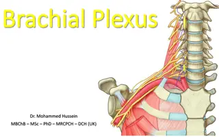
Differential Diagnosis Cervical disc herniation Carpal tunnel syndrome (median nerve at wrist) Cubital tunnel syndrome (ulnar nerve at elbow) Shoulder joint pain ( arthritis, adhesive capsulitis, trauma) Brachial plexitis (Parsonage Turner Syndrome) Pancoast syndrome and other neoplasms along the course of the brachial plexus Coronary artery disease, angina Thoracic outlet syndrome Spinal cord tumor/Syringomyelia ALS Central cord syndrome Complex regional pain syndrome Infection (bacterial, fungus, granuloma) Multiple sclerosis
Cervical disc herniation Etiology: Compression at neural foramen of one of the cervical nerve roots C5 T1 by disc herniation or bone spur. History: Neck pain, usually spontaneous, with radiation to ipsilateral scapula. Pain increases over time and radiates with paresthesias down the extremity in a dermatomal distribution. Pain is aggravated by neck movements, particularly with rotation toward the side of herniation. Muscular weakness may accompany the pain and paresthesias. Physical exam: The symptoms are usually reproduced by having the patient look up and toward the side of the pain (positive foraminal sign, Spurling Test). Check for muscular weakness, depressed deep tendon reflexes, and decreased sensation. Try to determine, particularly on sensory exam, a dermatomal pattern, and which nerve root is involved.
Cervical disc herniation Diagnostic studies: Cervical spine X-rays and MR scan are the most helpful in establishing the correct diagnosis. Treatment: NSAIDS, Pain meds, muscle relaxants and a soft cervical collar for initial management. When symptoms are not improved, the next steps are a short trial of Prednisone, cervical traction, and on rare occasions, a selective nerve root block by a pain specialist. If all of the above fail to relieve pain, or if the patient has a significant neurological deficit, refer for surgery.
Carpal Tunnel Syndrome Etiology: Compression of median nerve as it passes under the transverse carpal ligament at the wrist. Usually idiopathic, but may also be related to repetitive motion, trauma, inflammation, pregnancy, hypothyroidism or any condition which results in narrowing the space for the median nerve as it transits the carpal tunnel. Symptoms: The classic symptom, which is almost always present, is pain and numbness involving the hand and arm when awakening in the morning. It usually follows the median nerve distribution, but may also involve the entire length of the extremity. The pain and numbness is usually relieved by shaking the hand and arm. The characteristic symptoms are aggravated by holding a newspaper or steering wheel of a car. Repetitive motions such as data entry, meat cutting, jack hammering are frequently implicated.
Carpal Tunnel Syndrome Physical exam: Inspection in advanced cases will show atrophy of the thenar eminence (primarily opponens pollicis muscle). Motor testing may show weakness of the opponens pollicis muscle (Have patient hold tips of thumb and little finger tightly together. With your index finger you can easily break through this circle). On sensory exam the patient will have decreased sensation to touch, and pin in the distribution of the median nerve. Tinel sign and Phalen test are usually positive. Diagnostic studies: A positive sensory nerve conduction study demonstrating delay at the wrist usually clinches the diagnosis. In difficult cases ultrasound and MR scan can be helpful. Treatment: NSAIDS, wrist splint at night (but may also be used during the day), rest, avoid repetitive actions causing the problem. Surgical decompression of the median nerve at the wrist.
Cubital tunnel syndrome (tardy ulnar palsy) Etiology: Entrapment of the ulnar nerve as it wraps behind the medial epicondyle of the humerus in the ulnar groove ( funny bone ). History: The first symptom is usually the spontaneous onset of intermittent tingling and numbness in the ulnar distribution. This is usually accompanied by mild weakness of the hand. There may be a history of injury to the elbow, remote or recent. Patients frequently complain of elbow pain at the ulnar groove. Physical exam: On muscle testing look for weakness of the ulnar innervated intrinsic hand muscles. The 1st dorsal interosseous and abductor digiti quinti muscles are the best to examine. Decreased sensation to light touch and pin are present in the ulnar distribution and almost always split the ring finger. A strongly positive Tinel sign is elicited at the ulnar groove.
Cubital tunnel syndrome (tardy ulnar palsy) Diagnostic studies: Nerve conduction studies show delay across the ulnar groove at the elbow. Treatment: Protect the elbow from trauma, particularly avoid leaning on the elbow. Elbow pads are helpful. If symptoms increase the cubital tunnel is easily decompressed surgically as an outpatient procedure.