The world of microbiology delves into the study of various microorganisms like bacteria, fungi, protozoa, algae, parasites, viruses, and nematodes. It explores the classification and identification of bacteria based on shape, cell wall structure, Gram stain, cellular respiration, and growth factors. Learn about the different shapes and arrangements of bacterial cells, along with key vocabulary in the field of microbiology. Discover the fascinating realm of microbes and their varied characteristics.
Please find below an Image/Link to download the presentation.
The content on the website is provided AS IS for your information and personal use only. It may not be sold, licensed, or shared on other websites without obtaining consent from the author. If you encounter any issues during the download, it is possible that the publisher has removed the file from their server.
You are allowed to download the files provided on this website for personal or commercial use, subject to the condition that they are used lawfully. All files are the property of their respective owners.
The content on the website is provided AS IS for your information and personal use only. It may not be sold, licensed, or shared on other websites without obtaining consent from the author.
Bacteriology: study of bacteria Mycology: study of fungi Protozoology: study of protozoa Phycology (or algology): study of algae Parasitology: study of parasites Immunology: study of the immune system Virology: study of viruses Nematology: study of the nematodes Microbiology: study of microbes
Bacteria: Classification and Identification Bergey s Manual of Systematic Bacteriology Classifies bacteria via evolutionary or genetic relationships. Bergey s Manual of Determinative Bacteriology Classifies bacteria by cell wall composition, morphology, biochemical tests, differential staining, etc.
Bacteria: Classification and Identification 1. Based on shape 2. cell wall structure and Gram stain 3. cellular respiration 4. Growth factors (Energy Source and Nutrient Source)
Classify based on Shape Shape Coccus spherical, round, or ovoid Bacillus rod-shaped Spirillum spiral-shaped Vibrio comma-shaped Spirillum bacterial classification
Some are: Round cocci Streptococci chains Staphlococci in groups or clusters Diplococci in pairs Micrococci Strep Staph
Some are: Rod bacillus Streptobacilli chains Staphlobacilli in groups or clusters Diplobacilli in pairs
VOCAB Enteric of, relating to, or occurring in the intestines Nosocomal - originating in a hospital
Classify based on Gram stain Gram positive or Gram negative Gram positive bacteria appear purple Gram negative bacteria release the first dye used and appear red from the second (counter) dye Knowing Gram positive or Gram negative helps prescribe appropriate antibiotic The stain is named for H. C. J. Gram, a Danish physician who invented it in 1884.
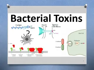
Gram Positives and Gram Negatives: Key Differences Gram positive have simpler, thicker walls, large amount of peptidoglycan Gram negative bacteria are thinner and have less peptidoglycan but more complex in structure An outer membrane on the Gram negative cell wall contains lipopolysaccharides (LPS) toxic substances responsible for making Gram negative organisms more threatening than Gram positives
Gram Positives Two layers: Outer cell wall - thick peptidoglycan layer, composed of complex cross- linked peptidoglycan, teichoic acid, polysaccharides and other proteins Cytoplasmic membrane - contains proteins that span the lipid bilayer Special Components of Gram Positive Bacteria Teichoic Acids anchor the outer cell wall to the cytoplasmic membrane by attaching to glycolipids also act as antigenic determinants (important for the serologic ID of Gram+ organisms) Polysaccharides
Examples of Gram Positive bacteria Streptococcus pyogenes - causes strep throat Staphylococcus aureus - causes skin infections and may be responsible for boils
Gram Negatives The Gram negative cell envelope has 3 layers (not including the periplasmic space): A unique outer membrane A thin peptidoglycan layer Cytoplasmic membrane Special Components of Gram negative Bacteria Outer membrane Complex, likely reason for difference in gram stain Murein lipoprotein starts in the peptidoglycan layer and extends outward to bind the unique third outer membrane. Lipopolysaccharide (LPS) major toxins of pathogenic Gram negative bacteria When the cell dies, LPS are released and can cause problems with organs or tissues
Classifying Bacteria by Cellular Respiration Aerobic bacteria, or strict aerobes - require oxygen Anaerobic bacteria, or strict anaerobes - cannot tolerate oxygen Facultative anaerobics are generally aerobes, but have the capacity to grow in the absence of oxygen Examples of Bacteria Classified by Cellular Respiration: Aerobic: Bacillus cereus Anaerobic: Clostridium spp. ( botulism, tetanus) Facultative anaerobes: Staphylococcus spp.
Classifying Bacteria by Growth Factors Under this scheme, they are generally classified according to: Energy source Nutrient source Energy Source Chemotroph chemical compounds as an energy source (most pathogenic bacteria are chemotrophs.) Phototroph - light as energy source Nutrient Source Heterotroph derive carbon from preformed organic nutrients such as sugar (most pathogenic bacteria are heterotrophs.) Autotroph derive carbon from inorganic sources such as carbon dioxide