Chronic Diarrhea and Malabsorption in Gastrointestinal Health
Explore the definitions, classifications, mechanisms, and clinical cases related to chronic diarrhea and malabsorption as discussed by Dr. Khalid Alswat, MD. Learn about the systematic approach to patients with these conditions and the next steps in diagnosis and management.

Download Presentation

Please find below an Image/Link to download the presentation.
The content on the website is provided AS IS for your information and personal use only. It may not be sold, licensed, or shared on other websites without obtaining consent from the author. If you encounter any issues during the download, it is possible that the publisher has removed the file from their server.
You are allowed to download the files provided on this website for personal or commercial use, subject to the condition that they are used lawfully. All files are the property of their respective owners.
The content on the website is provided AS IS for your information and personal use only. It may not be sold, licensed, or shared on other websites without obtaining consent from the author.
E N D
Presentation Transcript
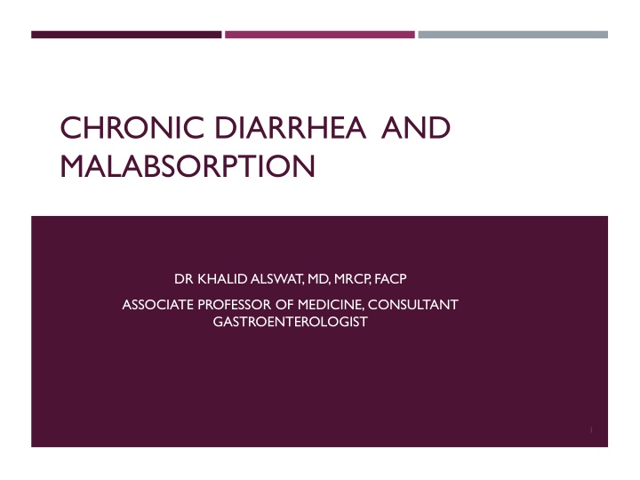
CHRONIC DIARRHEA AND MALABSORPTION DR KHALID ALSWAT, MD, MRCP, FACP ASSOCIATE PROFESSOR OF MEDICINE, CONSULTANT GASTROENTEROLOGIST 1
OBJECTIVES To recognize the definition and different classifications of chronic diarrhea To understand the mechanism of chronic diarrhea To learn systematic approach of patient with chronic diarrhea To understand the different mechanisms and causes of malabsorption To be able to recognize the clinical manifestation of malabsorption and approach to patient with malabsorption 2
CASE#1 Sarah is 22 Y F c/o non bloody diarrhea for 7 months 6-10 times/ Day Mild LIF pain Weight loss 10 kg ( 49 kg -> 39) O/E cachexia, LIF tenderness ( mild) Lab ( low hb 8 , low mcv) High ESR and CRP What is the most likely next STEP? 3
CASE#2 Ahmed is 18 years old with fatigue, abdominal bloating and diarrhea for 3 months. Itchy skin rash Unremarkable physical examination, except pallor and skin rash over thighs Has low Hb ( low mcv) Serology : anti TTG ab positive Most likely diagnosis and second step? 4
CASE #3 Nada is 27 years old F, with intermittent diffuse abd pain and diarrhea for 3 years. No blood in stool Diarrhea is triggered by fatty food, no nocturnal diarrhea Symptoms improved with defecation. Symptoms free for weeks Weight and appetite stable Examination: N CBC, ESR: N Most apropriate next step and diagnosis? 5
DEFINITIONS Diarrhea: decrease in fecal consistency (weight of stool and frequency are not reliable) Chronic diarrhea: > 4 weeks Diarrhea is a symptom, not a disease and may occur in many different conditions. Malabsorption: abnormality in absorption of food nutrients across the gastrointestinal (GI) tract. 6
DIARRHEA Acute diarrhea is a common and usually transient, self- limited, Infection related Chromic diarrhea: usually requires work up, non-infectious cause. 7
MECHANISM OF DIARRHEA Change in: Absorption Secretion Motility of the gut ->in response to various etiology 8
CLASSIFICATIONS Time course (acute vs. chronic) Volume (large vs. small) Pathophysiology (secretory vs. osmotic) Stool characteristics (watery vs. fatty vs. inflammatory). Epidemiology (epidemic vs. travel-related vs. immunosuppression-related) 9
OTHER CLASSIFICATION Fatty diarrhea Inflammatory diarrhea 11
COMMON CAUSES OF DIARRHEA Developing countries Developed countries Chronic bacterial Irritable bowel syndrome (IBS) Mycobacterial Inflammatory bowel disease (IBD) Parasitic infections Malabsorption syndromes (such as lactose intolerance and celiac disease) Then Functional disorders, Chronic infections (particularly in patients who are immunocompromised) Malabsorption Inflammatory bowel disease 12
CHRONIC DIARRHEA AND MALABSORPTION DR KHALID ALSWAT, MD, MRCP, FACP ASSOCIATE PROFESSOR OF MEDICINE, CONSULTANT GASTROENTEROLOGIST 13
APPROACH TO PATIENT WITH DIARRHEA History Physical examination Investigations Laboratory tests Radiology Endoscopy Management 14
HISTORY Main Symptom Consistency or frequency of stools, the presence of urgency or fecal soiling) Duration Acute or chronic ( weeks) Stool characteristics Greasy stools that float and are malodorous may suggest fat malabsorption while the presence of visible blood may suggest inflammatory bowel disease Volume of the diarrhea voluminous watery diarrhea is more likely to be due to a disorder in the small bowel while small-volume frequent diarrhea is more likely to be due to disorders of the colon Specific food Fatty , diary, allergy to certain food Change with fasting or night Secretory , IBS 15
HISTORY Specific food Fatty , diary, allergy to certain food Associated symptoms Extra-intestinal manifestation ( rash, arthritis. Mouth ulcers etc..) History of travel Drug hx Antibiotic use, others F Hx Of GI illnesses Systemic review ABD PAIN, Weight loss, appetite change Travelers diarrhea, parasitic etc e.g endocrine causes , thyroid, DM, Addison 16
PHYSICAL EXAMINATION Rarely provides a specific diagnosis. Findings suggestive of IBD (eg, mouth ulcers, a skin rash, episcleritis, an anal fissure or fistula, the presence of visible or occult blood on digital examination, abdominal masses or abdominal pain) Evidence of malabsorption (wasting, physical signs of anemia, scars indicating prior abdominal surgery) Lymphadenopathy (possibly suggesting, lymphoma, HIV infection) Palpation of the thyroid and examination for exophthalmos and lid retraction may provide support for a diagnosis of hyperthyroidism 17
INVESTIGATIONS ARE GUIDED BY HX AND PH EXAM (ESSENTIALS) CBC ESR Electrolytes Total protein and albumin TFT Stool : occult blood, C/S, ova and parasites, C-D toxin (if hx suggestive) 18
SPECIFIC INVESTIGATIONS The history and physical examination may point toward a specific diagnosis for which testing may be indicated 19
CLINICAL FEATURES Depend upon the cause and severity of the disease Malabsorption may either be global or partial (isolated). Global malabsorption: results from diseases associated with either diffuse mucosal involvement or a reduced absorptive surface An example is celiac disease in which diffuse mucosal disease can lead to impaired absorption of almost all nutrients Partial or isolated malabsorption: results from diseases that interfere with the absorption of specific nutrients. Defective cobalamin absorption, for example, can be seen in patients with pernicious anemia or those with disease (or resection) of the terminal ileum such as patients with Crohn's disease. 22
SIGNS AND SYMPTOMS OF MALABSORPTION Malabsorption of Clinical features Laboratory findings Calories Weight loss with normal appetite Pale and voluminous stool, diarrhea without flatulence, steatorrhea Fat Stool fat >6 g/day Hypoalbuminemia, hypoproteinemia Protein Edema, muscle atrophy, amenorrhea Watery diarrhea, flatulence, acidic stool pH, milk intolerance, stool osmotic gap Carbohydrates Increased breath hydrogen Anemia, subacute combined degeneration of the spinal cord (early symptoms are paresthesias and ataxia associated with loss of vibration and position sense) Macrocytic anemia, vitamin B12 decreased, abnormal Schilling test, serum methylmalonic acid and homocysteine increased Vitamin B12 23
SIGNS AND SYMPTOMS OF MALABSORPTION . Malabsorption of Clinical features Laboratory findings Macrocytic anemia, serum and RBC folate decreased, serum homocysteine increased Folic acid Anemia Cheilosis, painless glossitis, acrodermatitis, angular stomatitis Vitamin B, general Serum iron and ferritin decreased, total iron binding capacity increased Iron Microcytic anemia, glossitis, pagophagia Paresthesia, tetany, pathologic fractures due to osteomalacia, positive Chvostek and Trousseau signs Hypocalcemia, serum alkaline phosphatase increased, abnormal bone densitometry Calcium and vitamin D Vitamin A Follicular hyperkeratosis, night blindness Serum retinol decreased Prolonged prothrombin time, vitamin K-dependent coagulation factors decreased Vitamin K Hematoma, bleeding disorders 24
EXAMPLE OF MANIFESTATIONS ASSOCIATED WITH CELIAC DISEASE Dermatological manifestations Pale skin Neurological examination Motor weakness, peripheral neuropathy, or ataxia may be present . The Chvosteks sign or the Trousseau sign may be evident due to hypocalcemia . 25
INVESTIGATION FOR MALABSORPTION Investigations for nutrients deficiency Specific investigations to define the etiology and severity 26
EXAMPLES OF INVESTIGATIONS Common lab: CBC , ESR, CRP, PT, INR, LFT Serology: Anti TTG ( anti endomysial ab) Endoscopy Imaging 27
SEROLOGY No specific serology tests for all causes of malabsorption Serum Anti-TTG and antiendomysial antibodies can be used to help diagnose celiac sprue . Determination of fecal elastase and chymotrypsin can be used to try to distinguish between pancreatic causes and intestinal causes of malabsorption. 28
IAMGING Small bowel barium studies . Strictures Mucosal changes Diverticula CT scan of the abdomen Strictures Mucosal changes Diverticula Wall thickness Massesss, lymph nodes ERCP: pancreatitis (duct changes) , biliary doseases 29
IMAGING STUDIES Plain abdominal x-ray film: Pancreatic calcifications are indicative of chronic pancreatitis. 30
ENDOSCOPY Upper endoscopy with small bowel mucosal biopsy .Examples Celiac sprue Giardiasis Crohn disease Whipple disease Amyloidosis Lymphoma. Lower GI endoscopy: colonic and terminal ileal pathology ( e.g Chrons disease) 31
TREATMENT Treatment of causative diseases A gluten-free diet helps treat celiac disease . Similarly, a lactose-free diet Protease and lipase supplements are the therapy for pancreatic insufficiency . Antibiotics are the therapy for bacterial overgrowth . Corticosteroids, anti-inflammatory agents, such as mesalamine, and other therapies are used to treat CD 32
NUTRITIONAL SUPPORT Supplementing various minerals calcium, magnesium, iron, and vitamins Caloric and protein replacement also is essential . Medium-chain triglycerides can be used for lymphatic obstruction . In severe intestinal disease, such as massive resection and extensive regional enteritis, parenteral nutrition may become necessary 33
THANK YOU Additional essential reading .. 34
SECRETORY DIARRHEA CLUES: LARGE VOLUME ( >1 L/D); LITTLE CHANGE WITH FASTING; NORMAL OR LOW STOOL OSMOTIC GAP Bacterial toxins Disordered motility Inflammatory bowel disease Diabetic autonomic neuropathy o Crohn's disease o Irritable bowel syndrome o Ulcerative colitis o Postsympathectomy diarrhea o Microscopic colitis o Postvagotomy diarrhea o Collagenous colitis Ileal bile acid malabsorption Lymphocytic colitis Laxative abuse (stimulant laxatives) Vasculitis Medications and toxins Endocrinopathies Neoplasia Addison's disease o Colon carcinoma o Carcinoid syndrome o Lymphoma o Hyperthyroidism o Villous adenoma in rectum o Medullary carcinoma of the thyroid o Congenital syndromes (e.g., congenital chloridorrhea) Pheochromocytoma o Idiopathic 35 Gastrinoma, Somatostatinoma , VIPoma o
OSMOTIC DIARRHEA CLUES: STOOL VOLUME DECREASES WITH FASTING INCREASED STOOL OSMOTIC GAP 36
INFLAMMATORY DIARRHEA Inflammatory bowel disease Ulcerative colitis Crohn's disease Infectious diseases o o Invasive bacterial infections (e.g., tuberculosis, yersiniosis) o Invasive parasitic infections (e.g., amebiasis, strongyloidiasis) o Pseudomembranous colitis (Clostridium difficile infection) o Ulcerating viral infections (e.g., cytomegalovirus, herpes simplex virus) Ischemic colitis Radiation colitis Neoplasia Colon cancer Lymphoma o o 37 o
FATTY DIARRHEA Malabsorption syndromes o Mucosal diseases o Short bowel syndrome Postresection diarrhea o Small bowel bacterial overgrowth o Mesenteric ischemia Maldigestion: o Pancreatic exocrine insufficiency o Inadequate luminal bile acid 38
PHASES OF ABSORPTION Luminal phase: dietary fats, proteins, and carbohydrates are hydrolyzed and solubilized by secreted digestive enzymes and bile. Mucosal phase: relies on the integrity of the brush-border membrane of intestinal epithelial cells to transport digested products from the lumen into the cells. Postabsorptive phase: nutrients are transported via lymphatics and portal circulation from epithelial cells to other parts of the body. 39
LUMINAL PHASE Phase and nature of malabsorptive defect A. Substrate hydrolysis 1. Digestive enzyme deficiency 2. Digestive enzyme inactivation 3. Dyssynchrony of enzyme release, inadequate mixing B. Fat solubilization 1. Diminished bile salt synthesis 2. Impaired bile secretion 3. Bile salt de-conjugation 4. Increased bile salt loss C. Luminal availability of specific nutrients 1. Diminished gastric acid 2. Diminished intrinsic factor Example Chronic pancreatitis Zollinger-Ellison syndrome Post Billroth II procedure Cirrhosis Chronic cholestasis Bacterial overgrowth Ileal disease or resection Atrophic gastritis - vitamin B12 Pernicious anemia - vitamin B12 40 3. Bacterial consumption of nutrients Bacterial overgrowth - vitamin B12
MUCOSAL (ABSORPTIVE) PHASE Phase and nature of malabsorptive defect A. Brush border hydrolysis* 1. Congenital disaccharidase defect 2. Acquired disaccharidase defect B. Epithelial transport 1. Nutrient-specific defects in transport 2. Global defects in transport (a) decreased absorptive surface area Example Sucrase-isomaltase deficiency Lactase deficiency Hartnup's disease Celiac sprue intestinal resection celiac sprue, tropical sprue, giardiasis, Crohn disease, AIDS enteropathy, chemotherapy, or radiation therapy; lymphoma and amyloidosis (b) damaged absorbing surface (c) infiltrating disease of the intestinal wall 41 * This process is sometimes considered as part of the luminal phase.
POSTABSORPTIVE, PROCESSING PHASE Phase and nature of malabsorptive defect A. Enterocyte processing Example Abetalipoproteinemia Both congenital (e.g. intestinal lymphangiectasia) Acquired (e.g. Whipple diseases , lymphoma, tuberculosis), B. Obstruction of Lymphatic 42
LAB STUDIES Hematological tests A CBC Serum iron, vitamin B-12, and folate Prothrombin time. Electrolytes and chemistries Hypokalemia, hypocalcemia, hypomagnesemia, and metabolic acidosis . Protein malabsorption may cause hypoproteinemia and hypoalbuminemia . Fat malabsorption can lead to low serum levels of triglycerides, cholesterol ESR which is elevated in inflammatory diseases like Crohn disease and Whipple disease 43
STOOL ANALYSIS Stool pH may be assessed. Values of <5.6 are consistent with carbohydrate malabsorption Stool C/S Pus cells in the stool e.g IBD, some infections 44
TESTS OF FAT MALABSORPTION For a quantitative measurement of fat absorption, a 72-hour fecal fat collection Qualitative test Sudan III stain of stool, less reliable . 45
Stool fat pancreatic, celiac inflammatory -- IBD ,infection 46
SCHILLING TEST Malabsorption of vitamin B-12 may occur as a consequence of: Deficiency of intrinsic factor (eg, pernicious anemia, gastric resection) Pancreatic insufficiency, bacterial overgrowth Ileal resection, or disease . 48
3 STEPS SCHILLING TEST 1. Oral VIT B12 2. VIT B12 orally +intrinsic factor 3. VIT B12 orally +intrinsic factor+ oral antibiotics 49
BACTERIAL OVERGROWTH Bacterial overgrowth cause an early rise in breath hydrogen JEUJENAL CULTURE 14c D xylose breath test ,high sensitivity and specificity 50


 PHASE")