Explore the intricate workings of the circulatory and respiratory systems, from the alveoli in the lungs to the major arteries and veins that transport blood throughout the body. Learn about key components like the diaphragm, bronchi, and cardiac cycle, essential for oxygen exchange and nutrient delivery. Gain insights into closed and open circulatory systems and the importance of pulmonary circulation in oxygenation.
Please find below an Image/Link to download the presentation.
The content on the website is provided AS IS for your information and personal use only. It may not be sold, licensed, or shared on other websites without obtaining consent from the author. If you encounter any issues during the download, it is possible that the publisher has removed the file from their server.
You are allowed to download the files provided on this website for personal or commercial use, subject to the condition that they are used lawfully. All files are the property of their respective owners.
The content on the website is provided AS IS for your information and personal use only. It may not be sold, licensed, or shared on other websites without obtaining consent from the author.
Glossary alveolus: (plural: alveoli) (also, air sacs) the terminal structure of the lung passage where gas exchange occurs aorta: the major artery that takes blood away from the heart to the systemic circulatory system artery: a blood vessel that takes blood away from the heart atrium: (plural: atria) a chamber of the heart that receives blood from the veins bicuspid valve: a one-way opening between the atrium and the ventricle in the left side of the heart capillary: the smallest blood vessel that allows the passage of individual blood cells and the site of diffusion of oxygen and nutrient exchange cardiac cycle: the filling and emptying the heart of blood caused by electrical signals that cause the heart muscles to contract and relax
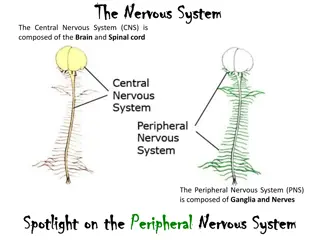
closed circulatory system: a system that has the blood separated from the bodily interstitial fluid and contained in blood vessels diaphragm: a skeletal muscle located under lungs that encloses the lungs in the thorax diastole: the relaxation phase of the cardiac cycle when the heart is relaxed and the ventricles are filling with blood electrocardiogram (ECG): a recording of the electrical impulses of the cardiac muscle inferior vena cava: the major vein of the body returning blood from the lower parts of the body to the right atrium open circulatory system: a circulatory system that has the blood mixed with interstitial fluid in the body cavity and directly bathes the organs
bronchi: (singular: bronchus) smaller branches of cartilaginous tissue that stem off of the trachea; air is funneled through the bronchi to the region where gas exchange occurs in the alveoli bronchiole: an airway that extends from the main bronchus to the alveolar sac larynx: the voice box, located within the throat nasal cavity: an opening of the respiratory system to the outside environment pharynx: the throat primary bronchus: (also, main bronchus) a region of the airway within the lung that attaches to the trachea and bifurcates to form the bronchioles pulmonary circulation: the flow of blood away from the heart through the lungs where oxygenation occurs and then back to the heart superior vena cava: the major vein of the body returning blood from the upper part of the body to the right atrium
systemic circulation: the flow of blood away from the heart to the brain, liver, kidneys, stomach, and other organs, the limbs, and the muscles of the body, and then back to the heart systole: the contraction phase of cardiac cycle when the ventricles are pumping blood into the arteries trachea: the cartilaginous tube that transports air from the throat to the lungs tricuspid valve: a one-way opening between the atrium and the ventricle in the right side of the heart vein: a blood vessel that brings blood back to the heart ventricle: (of the heart) a large chamber of the heart that pumps blood into arteries
Animals are complex multicellular organisms that require a mechanism for transporting nutrients throughout their bodies and removing wastes. The human circulatory system has a complex network of blood vessels that reach all parts of the body. This extensive network supplies the cells, tissues and organs with oxygen and nutrients and removes carbon dioxide and waste compounds. The medium for transport of gases and other molecules is the blood, which continually circulates through the system. Pressure differences within the system cause the movement of the blood and are created by the pumping of the heart.
Gas exchange between tissues and the blood is an essential function of the circulatory system. In humans, other mammals, and birds, blood absorbs oxygen and releases carbon dioxide in the lungs. Thus the circulatory and respiratory system, whose function is to obtain oxygen and discharge carbon dioxide, work in tandem.
The primary function of the respiratory system is to deliver oxygen to the cells of the body s tissues and remove carbon dioxide, a cell waste product. The main structures of the respiratory system are the nasal cavity, the trachea, and lungs. All aerobic organisms require oxygen to carry out their metabolic functions. Along the evolutionary tree, different organisms have devised different means of obtaining oxygen from the surrounding atmosphere. The environment in which the animal lives greatly determines how an animal respires. The complexity of the respiratory system is correlated with the size of the organism. As animal size increases, diffusion distances increase and the ratio of surface area to volume drops. In unicellular organisms, diffusion across the cell membrane is sufficient for supplying oxygen to the cell.
Diffusion is a slow, passive transport process. Diffusion means a feasible way of providing oxygen to the cell, the rate of oxygen uptake must match the rate of diffusion across the membrane. In other words, if the cell were very large or thick, diffusion would not be able to provide oxygen quickly enough to the inside of the cell. Therefore, dependence on diffusion as a means of obtaining oxygen and removing carbon dioxide remains feasible only for small organisms or those with highly-flattened bodies, such as many flatworms (Platyhelminthes). Larger organisms had to evolve specialized respiratory tissues, such as gills, lungs, and respiratory passages accompanied by a complex circulatory systems, to transport oxygen throughout their entire body.
Direct Diffusion For small multicellular organisms, diffusion across the outer membrane is sufficient to meet their oxygen needs. Gas exchange by direct diffusion across surface membranes is efficient for organisms less than 1 mm in diameter. In simple organisms, such as cnidarians and flatworms, every cell in the body is close to the external environment. Their cells are kept moist and gases diffuse quickly via direct diffusion. Flatworms are small, literally flat worms, which breathe through diffusion across the outer membrane. The flat shape of these organisms increases the surface area for diffusion, ensuring that each cell within the body is close to the outer membrane surface and has access to oxygen. If the flatworm had a cylindrical body, then the cells in the center would not be able to get oxygen.
In mammals, pulmonary ventilation occurs via inhalation (breathing). During inhalation, air enters the body through the nasal cavity located just inside the nose. As air passes through the nasal cavity, the air is warmed to body temperature and humidified. The respiratory tract is coated with mucus to seal the tissues from direct contact with air. Mucus is high in water. As air crosses these surfaces of the mucous membranes, it picks up water. These processes help equilibrate the air to the body conditions, reducing any damage that cold, dry air can cause. Particulate matter that is floating in the air is removed in the nasal passages via mucus and cilia. The processes of warming, humidifying, and removing particles are important protective mechanisms that prevent damage to the trachea and lungs. Thus, inhalation serves several purposes in addition to bringing oxygen into the respiratory system.
Terminal bronchioles are connected by respiratory bronchioles to alveolar ducts and alveolar sacs. Each alveolar sac contains 20 to 30 spherical alveoli and has the appearance of a bunch of grapes. Air flows into the atrium of the alveolar sac, then circulates into alveoli where gas exchange occurs with the capillaries. Mucous glands secrete mucous into the airways, keeping them moist and flexible.
Protective Mechanisms The air that organisms breathe contains particulate matter such as dust, dirt, viral particles, and bacteria that can damage the lungs or trigger allergic immune responses. The respiratory system contains several protective mechanisms to avoid problems or tissue damage. In the nasal cavity, hairs and mucus trap small particles, viruses, bacteria, dust and dirt to prevent their entry. If particulates do make it beyond the nose, or enter through the mouth, the bronchi and bronchioles of the lungs also contain several protective devices. The lungs produce mucus a sticky substance made of mucin, a complex glycoprotein, as well as salts and water that traps particulates. The bronchi and bronchioles contain cilia, small hair-like projections that line the walls of the bronchi and bronchioles. These cilia beat in unison and move mucus and particles out of the bronchi and bronchioles back up to the throat where it is swallowed and eliminated via the esophagus.
Summary Animal respiratory systems are designed to facilitate gas exchange. In mammals, air is warmed and humidified in the nasal cavity. Air then travels down the pharynx, through the trachea, and into the lungs. In the lungs, air passes through the branching bronchi, reaching the respiratory bronchioles, which house the first site of gas exchange. The respiratory bronchioles open into the alveolar ducts, alveolar sacs, and alveoli. Because there are so many alveoli and alveolar sacs in the lung, the surface area for gas exchange is very large. Several protective mechanisms are in place to prevent damage or infection. These include the hair and mucus in the nasal cavity that trap dust, dirt and other particulate matter before they can enter the system. In the lungs, particles are trapped in a mucus layer and transported via cilia up to the esophageal opening at the top of the trachea to be swallowed.
The Circulatory System The circulatory system is a network of vessels the arteries, veins, and capillaries and a pump, the heart. In all vertebrate organisms this is a closed- loop system, in which the blood is largely separated from the body s other compartment, the interstitial fluid, which is the fluid bathing the cells. Blood circulates inside blood vessels and circulates unidirectional from the heart around one of two circulatory routes, then returns to the heart again; this is a closed circulatory system. Open circulatory systems are found in invertebrate animals in which the circulatory fluid bathes the internal organs directly even though it may be moved about with a pumping heart. extracellular fluid
The Cardiac Cycle The main purpose of the heart is to pump blood through the body; it does so in a repeating sequence called the cardiac cycle. The cardiac cycle is the flow of blood through the heart coordinated by electrochemical signals that cause the heart muscle to contract and relax. In each cardiac cycle, a sequence of contractions pushes out the blood, pumping it through the body; this is followed by a relaxation phase, where the heart fills with blood. These two phases are called the systole (contraction) and diastole (relaxation), respectively. The signal for contraction begins at a location on the outside of the right atrium. The electrochemical signal moves from there across the atria causing them to contract.
The contraction of the atria forces blood through the valves into the ventricles. Closing of these valves caused by the contraction of the ventricles produces a lub sound. The signal has, by this time, passed down the walls of the heart, through a point between the right atrium and right ventricle. The signal then causes the ventricles to contract. The ventricles contract together forcing blood into the aorta and the pulmonary arteries. Closing of the valves to these arteries caused by blood being drawn back toward the heart during ventricular relaxation produces a dub sound.
In each cardiac cycle, a series of contractions (systoles) and relaxations (diastoles) pumps blood through the heart and through the body. (a) During cardiac diastole, blood flows into the heart while all chambers are relaxed. (b) Then the ventricles remain relaxed while atrial systole pushes blood into the ventricles. (c) Once the atria relax again, ventricle systole pushes blood out of the heart.
The pumping of the heart is a function of the cardiac muscle cells, or cardiomyocytes, that make up the heart muscle. Cardiomyocytes are distinctive muscle cells that are striated like skeletal muscle but pump rhythmically and involuntarily like smooth muscle; adjacent cells are connected by intercalated disks found only in cardiac muscle. These connections allow the electrical signal to travel directly to neighboring muscle cells. The electrical impulses in the heart produce electrical currents that flow through the body and can be measured on the skin using electrodes. This information can be electrocardiogram (ECG) a recording of the electrical impulses of the cardiac muscle. observed as an
Blood Vessels The blood from the heart is carried through the body by a complex network of blood vessels. Arteries take blood away from the heart. The main artery of the systemic circulation is the aorta; it branches into major arteries that take blood to different limbs and organs. The aorta and arteries near the heart have heavy but elastic walls that respond to and smooth out the pressure differences caused by the beating heart. Arteries farther away from the heart have more muscle tissue in their walls that can constrict to affect flow rates of blood. The major arteries diverge into minor arteries, and then smaller vessels called arterioles, to reach more deeply into the muscles and organs of the body.
Arterioles diverge into capillary beds. Capillary beds contain a large number, 10 s to 100 s of capillaries that branch among the cells of the body. Capillaries are narrow-diameter tubes that can fit single red blood cells and are the sites for the exchange of nutrients, waste and oxygen with tissues at the cellular level. Fluid also leaks from the blood into the interstitial space from the capillaries. The capillaries converge again into venules that connect to minor veins that finally connect to major veins. Veins are blood vessels that bring blood high in carbon dioxide back to the heart. Veins are not as thick-walled as arteries, since pressure is lower, and they have valves along their length that prevent backflow of blood away from the heart. The major veins drain blood from the same organs and limbs that the major arteries supply.
I. Circulatory System A. Main functions: circulation of heat (yes heat!), nutrients, hormones, and gases B. Two major types 1. Open a. insects have open circulation - blood not all in vessels, but sloshes around body cavity b. works okay for small organisms, but is inefficient for distributing materials, especially oxygen, in large organisms 2. Closed a. all higher animals have closed systems, including earthworms b. more efficient C. We'll concentrate on closed systems D. See handout on how heart works to circulate blood in humans E. Terminology 1. Arteries - blood vessels that carry blood away from heart 2. Veins - vessels that carry blood to heart 3. Capillaries - very small blood vessels where most gas exchange occurs 4. Atria - chambers in heart that accept blood from veins 5. Ventricles - chambers that push blood out of heart F. Evolution of Circulatory Systems 1. Fish - two chambered heart 2. Amphibians and Reptiles - three chambered heart 3. Humans and Birds - four chambered heart
G. Heart Function in Humans 1. Two phases for each heartbeat (normal heart beat takes 0.8 sec) a. systolic - when heart contracts i. takes 0.1 sec for atrium to contract ii. takes 0.3 sec for ventricle to contract iii. total systolic time is 0.4 sec b. diastolic - when heart is relaxed between beats i. 0.7 sec for atria to fill back up (starts right after contraction) ii. 0.4 sec for ventricles to fill back up c. typical heartrate is 75 beats/min, but can vary greatly depending on level of activity 2. Valves help control flow of blood into and out of heart a. atrioventricular - keeps blood from flowing back into atria when ventricles contract - located between atria and ventricles b. semilunar - keeps blood from going back into ventricles when heart is diastolic, located at exit of ventricles c. sinoatrial node - electrical nerve center -sends electrical signal that induces heart muscle to contract 3. Cardiac Output - example for average person a. 75 beats/min b. 70 mls/beat c. 5.25 liters/min - about entire blood supply circulated once a minute 4. Heartrate inversely proportional to body size -slowest in elephants, fastest in hummingbirds
5. Heart rate in humans depends on temperature (goes up with temp), and on hormones (adrenaline stimulates heartrate), and carbon dioxide in blood (too much lowers pH, carbonic acid, and heart rate picks up) 6. Blood flows fastest right out of heart, slowest in capillaries. Blood pressure highest at exit from heart, lowest in veins II. Respiration A. Primitive organisms can "breathe" through their skin - but must be small and flat to get enough surface area to do this. Also, skin needs to be moist, or exchange is too slow B. Other organisms use gills or lungs 1. Fish - use gills. Work well in water, but would be hard to keep intact in dry air (wind, dessication, etc.) 2. Terrestrial organisms use lungs, although insects do not have lungs 3. Insects have trachea (tubes that go all through their body). Openings on their body are called spiracles, which let oxygen in and carbon dioxide out 4. In mammals, as chest expands, it draws out the lungs, creating negative pressure, and air rushes into lungs. We do not "push" air into our lungs, but frogs do 5. Air enters trachea (windpipe) and goes into large tubes called bronchi. 6. Bronchi branch into bronchioles, smaller tubes, and eventually end at the alveoli, small sacs where gas exchange occurs in lungs 7. Mucus and cilia line the bronchi and bronchioles - keep dust particles out 8. Each breath brings in about 500 ml (1/2 liter) of air 9. Stretch sensors in lungs determine when a breath ends 10. Breathing rate - controlled by blood pH (too low, more rapid breathing)
11. Exchange of gases in lungs i. partial pressure of oxygen is greatest in atmosphere (21%, or 210,000 ppm) ii. is much lower in lungs, hence oxygen diffuses into lungs iii. partial pressure of carbon dioxide is much higher in lungs than in atmosphere (20,000 ppm vs 365 ppm) and hence carbon dioxide diffuses out of lungs, not in! 12. Hemoglobin is used to facilitate movement of oxygen in blood. Each molecule can bind 4 oxygens 13. Each hemoglobin molecule contains 4 iron molecules, which are where the oxygen molecules bind 14. Some insects have hemocyanin, which has copper instead of iron makes their blood bluish instead of reddish C. Lung Diseases 1. Asthma - involuntary constrictions of the bronchioles, leading to breathing distress. Is on the rise among urban children, for reasons unknown 2. Asthma treated by giving sprays with histamines - causes tubes to relax and open (epinephrine for example) 3. Black Lung - accumulation of coal dust in lungs - common among coal miners but not as prevalent today as in the past. Devastating disease 4. Particulate inhalation a major concern today - diesel engine particles
Summary Animal respiratory systems are designed to facilitate gas exchange. In mammals, air is warmed and humidified in the nasal cavity. Air then travels down the pharynx and larynx, through the trachea, and into the lungs. In the lungs, air passes through the branching bronchi, reaching the respiratory bronchioles. The respiratory bronchioles open up into the alveolar ducts, alveolar sacs, and alveoli. Because there are so many alveoli and alveolar sacs in the lung, the surface area for gas exchange is very large. The mammalian circulatory system is a closed system with double circulation passing through the lungs and the body. It consists of a network of vessels containing blood that circulates because of pressure differences generated by the heart.
The heart contains two pumps that move blood through the pulmonary and systemic circulations. There is one atrium and one ventricle on the right side and one atrium and one ventricle on the left side. The pumping of the heart is a function of cardiomyocytes, distinctive muscle cells that are striated like skeletal muscle but pump rhythmically and involuntarily like smooth muscle. The signal for contraction begins in the wall of the right atrium. The electrochemical signal causes the two atria to contract in unison; then the signal causes the ventricles to contract. The blood from the heart is carried through the body by a complex network of blood vessels; arteries take blood away from the heart, and veins bring blood back to the heart.
Exercises Which of the following statements about the human respiratory system is false? When we breathe in, air travels from the pharynx to the trachea. The bronchioles branch into bronchi. Alveolar ducts connect to alveolar sacs. Gas exchange between the lungs and blood takes place in the alveolus. Which of the following statements about the circulatory system is false? Blood in the pulmonary vein is deoxygenated. Blood in the inferior vena cava is deoxygenated. Blood in the pulmonary artery is deoxygenated. Blood in the aorta is oxygenated. The respiratory system ________. provides body tissues with oxygen provides body tissues with oxygen and carbon dioxide establishes how many breaths are taken per minute provides the body with carbon dioxide Which is the order of airflow during inhalation? nasal cavity, trachea, larynx, bronchi, bronchioles, alveoli nasal cavity, larynx, trachea, bronchi, bronchioles, alveoli nasal cavity, larynx, trachea, bronchioles, bronchi, alveoli nasal cavity, trachea, larynx, bronchi, bronchioles, alveoli
Where does the right ventricle send blood? the head the upper body the lungs the lower body During the systolic phase of the cardiac cycle, the heart is ________. contracting relaxing contracting and relaxing filling with blood How do arteries differ from veins? Arteries have thicker wall layers to accommodate the changes in pressure from the heart. Arteries carry blood. Arteries have thinner wall layers and valves and move blood by the action of skeletal muscle. Arteries are thin walled and are used for gas exchange. Describe the function of these terms and describe where they are located: main bronchus, trachea, alveoli. How does the structure of alveoli maximize gas exchange? Describe the cardiac cycle.
Answers B A A B C A A The main bronchus is the conduit in the lung that funnels air to the airways where gas exchange occurs. The main bronchus attaches the lungs to the very end of the trachea where it bifurcates. The trachea is the cartilaginous structure that extends from the pharynx to the lungs. It serves to funnel air to the lungs. The alveoli are the site of gas exchange; they are located at the terminal regions of the lung and are attached to the alveolar sacs, which come from the alveolar ducts and respiratory bronchioles terminal bronchi. The sac-like structure of the alveoli increases their surface area. In addition, the alveoli are made of thin-walled cells. These features allows gases to easily diffuse across the cells. The heart receives an electrical signal triggering the cardiac muscle cells in the atria to contract. The signal pauses before passing onto the ventricles so the blood is pumped through the body. This is the systolic phase. The heart then relaxes in diastole and fills again with blood.


 smaller branches of")


")