Clinical Importance of Anaerobes in Microbiology Lectures
Explore the significance of anaerobic bacteria, their classification, habitats, and clinical relevance in human infections. Learn about anaerobic culture, identification methods, and treatment approaches for anaerobic-associated diseases.

Download Presentation

Please find below an Image/Link to download the presentation.
The content on the website is provided AS IS for your information and personal use only. It may not be sold, licensed, or shared on other websites without obtaining consent from the author. If you encounter any issues during the download, it is possible that the publisher has removed the file from their server.
You are allowed to download the files provided on this website for personal or commercial use, subject to the condition that they are used lawfully. All files are the property of their respective owners.
The content on the website is provided AS IS for your information and personal use only. It may not be sold, licensed, or shared on other websites without obtaining consent from the author.
E N D
Presentation Transcript
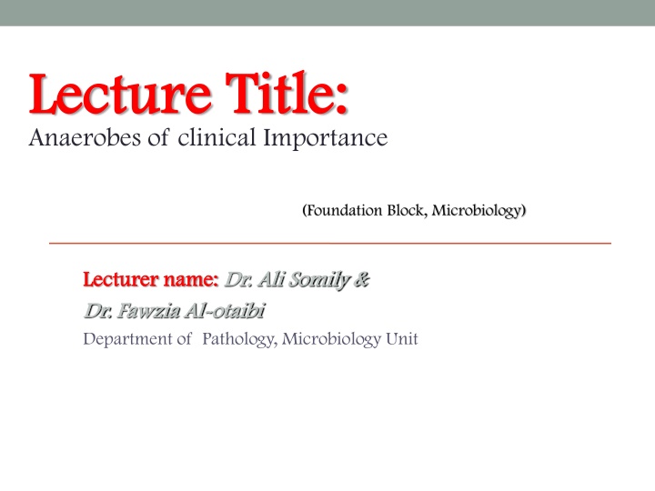
Lecture Title: Lecture Title: Anaerobes of clinical Importance (Foundation Block, Microbiology) Lecturer name: Lecturer name: Dr. Ali Somily & Dr. Ali Somily & Dr. Dr. Fawzia Fawzia Al Al- -otaibi otaibi Department of Pathology, Microbiology Unit
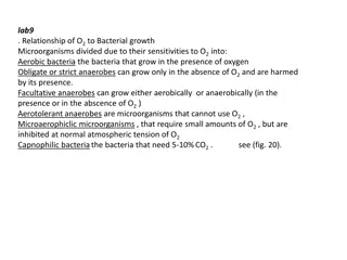
LECTURE OBJECTIVES By the end of this lecture the student should be able to: By the end of this lecture the student should be able to: Describe anaerobic bacteria including their sensitivity to oxygen and where they may be found in the environment and the human body. Differentiate the various types of anaerobes with regard to atmospheric requirement (i.e. obligate anaerobes, Faculative anaerobes and aerotolerent anaerobes. Describe how anaerobes, as part of endogenous microbiota, initiate and establish infection. Name the endogenous anaerobes commonly involved in human infection.
LECTURE OBJECTIVES Recognize specimens that are acceptable and unacceptable for anaerobic culture. Give the clues(sign and manifestations) to anaerobic infection, name the most probable etiologic agents of the following(Wound botulism, gas gangrene,tetanus,Actinomycosis,Pseudomembranous colitis and bacterial vaginosis) Describe the microscopic and colony morphology and the results of differentiating anaerobic isolates. Discuss antimicrobial susceptibility testing of anaerobes including methods and antimicrobial agents to be tested. Describe the major approaches to treat anaerobic-associated diseases either medical or surgical.
CLASSIFICATION CLASSIFICATION Anaerobic spore forming bacilli (Clostridia) Gram negative bacilli non-sporing forming (Bacteroides) Anaerobic streptococci (Peptostreptococcus) Anaerobic staphylococcus (Peptococcus) Gram negative diplococci (Veillonella) Gram positive bacilli (Actinomyces)
ANAEROBIOSIS ANAEROBIOSIS Lack cytochrome-cannot use oxygen as hydrogen acceptor Most Lack Catalase & Peroxidase Contain flavoprotein so in the presence of oxygen produce H2O2 which is toxic Some lack enzyme superoxide dismutase so many killed , peroxide and toxic radicles enzyme like fumarate reductase must be in reduced form to work
HABITAT I : HABITAT I : These organism are normal flora in: A. Oropharynx eg. 1. Provetella melaninogenicus 2. Fusobacteria 3. Veillonella B. Gastrointestinal tract Found mainly in the large colon in large numbers Total number of anaerobes = 10 11 While all aerobes (including E. coli) = 10 14 examples are (1) B acteroides fragilis (2) Bifidobacterium species C. Female genital tract (mainly in the vagina)
FEATURES OF ANAEROBIC INFECTIONS FEATURES OF ANAEROBIC INFECTIONS Infections Infections are always near to the site of the body which are are always near to the site of the body which are habitat. habitat. Infection from animal bites. 1. Deep abscesses 2. The infections are also polymicrobial 3. Gas formation, foul smell 4. Detection of "Sulphur granules"' due to actinomycosis 5. Failure to grow organism from pus if not culture anaerobically. 6. Failure to respond to usual antibiotics. 7.
HOW DOES THE INFECTION BEGIN ? DISRUPTION OF BARRIERS TRAUMA OPERATIONS CANCEROUS INVASION OF TISSUES DISRUPTION OF BLOOD SUPPLY DROPS OXYGEN CONTENT OF TISSUE DECREASE IN Eh POTENTIAL TISSUE NECROSIS
WHAT ARE THE INFECTION CAUSED BY WHAT ARE THE INFECTION CAUSED BY THESE ANAEROBIC ORGANISMS I THESE ANAEROBIC ORGANISMS I Post operative wound infection Brain, dental, lung abscess Intra abdominal abscess, appendicitis, diverculitis Infection of the female genital tract: Septic abortion, puerperal infection and endometritis , pelvic abscess or breast abscess Diabetic foot infections and pilonidal sinus
LABORATORY DIAGNOSIS: LABORATORY DIAGNOSIS: When anaerobic infection is suspected; a) Specimens have to be collected from the site containing necrotic tissue. b) Pus is better than swabs. c) Specimens has to be send to the laboratory within 1/2 hour why? d) Fluid media like cooked meat broth are the best culture media. e) Specimens have to incubated anaerobically for 48 hours.
TREATMENT: TREATMENT: Bacteroides fragilis is always resistant to penicillin. But penicillin can he used for other anaerobes Flagyl (metronidazole) is the drug of choice. Clindamycin can also be used.
ANAEROBIC NON SPORE FORMING BACILLI ACTINOMYCES SPP ACTINOMYCOSIS PROPIONIBACTERIUM SPP ACNE MOBILUNCUS SPP BACTERIAL VAGINOSIS LACTOBACILLUS SPP ENDOCARDITIS EUBACTERIUM SPP BIFIDOBACTERIUM SPP
ACTINOMYCOSIS Actinomyces are branching anaerobic or microaerophilic Gram positive bacilli Source of the infection is normal flora and the host usually normal host Primary site of the infection is mouth, lung, appendix, uterus with IUD (chronic infection) Infection can spread to the brain, liver, bone and blood Diagnosis by Gram stain with sulfur granules and growth of molar tooth colonies Treatment penicillin, clindamycin or tetracycline
ORGANISM GROUPS GRAM NEGATIVE RODS BACTEROIDES PREVOTELLA PORPHYROMONAS FUSOBACTERIUM BUTYRIVIBRIO SUCCINOMONAS
BACTEROIDES BACTEROIDES STRICT ANAEROBE PLEOMORPHIC GRAM NEGATIVE BACILLI (COCCO BACILLI) NORMAL FLORA IN OROPHARYNX GASTROINTESTINAL TRACT VAGINA
BACTEROIDES BACTEROIDES GROUP = B. FRAGILIS, B. VULGARIS, B.THETAIOTAMICRON, B. UNIFORMIS ACCOUNT FOR 1/3 OF ALL ISOLATES RESISTANT TO 20% BILE RESISTANT TO MANY ANTIBIOTICS PENICILLIN, KANAMYCIN, VANCOMYCIN, COLISTIN AND MANY MORE NO PIGMENTATION OF COLONIES OR FLUORESCENCE
BACTEROIDES OTHER SP BACTEROIDES OTHER SP BACTEROIDES SPECIES OTHER THAN B. FRAGILIS GROUP BILE SENSITIVE RESISTANT TO KANAMYCIN ONLY SOME PIGMENTED
OTHER GRAM NEGATIVE RODS FUSOBACTERIUM NECROPHORUM FUSOBACTERIUM NECROPHORUM GRAM NEGATIVE BACILLI PERITONISILLAR INTRNAL JUGULAR VEIN THROMBOSIS EMBOLI TO THE LUNG PEPTOCOCCUS PEPTOCOCCUS GRAM POSITIVE COCCI IN CLUSTERS PEPTOSTREPTOCOCCUS PEPTOSTREPTOCOCCUS GRAM POSITIVE COCCI IN CHAINS BRAIN ABSCESS VEILLONELLA PARVULA VEILLONELLA PARVULA GRAM NEGATIVE COCCI
CLOSTRIDIUM SPECIES CLOSTRIDIUM SPECIES LARGE GRAM POSITIVE RODS SPORE FORMATION Causative Agents For 1.Gas gangrene : Cl. perfringens and other e.g septicum 2.Tetanus : Cl. tetani 3.Botulism : Cl. botulinum 4.Toxic enterocolitis : Cl. difficile (Pseudomembernous colitis)
Clostridium Clostridium perfringens perfringens (CI . (CI . welchii welchii) ) Morphology large rods gram +ve with bulging endospores Laboratory diagnosis Smear Gram stain Large Gram positive bacilli with few or no WBCs Culture Blood agar with haemolytic colonies (double zone of haemolysis ) Cooked meat medium Gives the NAGLAR'S Reaction & toxin neutralization on Egg yolk medium & toxin is a phospholipase
Clostridium Clostridium perfringens perfringens (CI . (CI . welchii welchii) ) Can leads to the following diseases 1) Wound Contamination 2) Wound infection 3) Gas Gangrene Gas Gangrene - most important disease 4) Gas Gangrene of the uterus in criminal abortion 5) Food Poisoning : Spores are swallowed Germinate in gut after 18 hours(Toxin production) abdominal pain and diarrhoea
Clostridium Clostridium perfringens perfringens (CI . (CI . welchii welchii) ) Pathogenesis: Traumatic open wounds or compound fractures lead to muscle damages and contamination with dirt etc, Mainly in war wounds, old age, low blood supply and amputation of thigh (required prophylaxis with penicillin Prevention and Treatment Remove dead tissue , debris and foreign bodies .Penicillin and hyperbaric oxygen in some cases
Cl.tetani Cl.tetani ( (TETANUS TETANUS) Morphology gram +ve anaerobic with terminal spore. Drum Stick appearance Lives in soil and animal feaces. e,g horse and any wound can infected if contaminated by spores Face & neck wounds are more dangerous
Cl.tetani Cl.tetani ( (TETANUS TETANUS) Clinical Features Incubation period 1-3 weeks (time from infection to the appearance of symptoms) Symptoms: local (not common), cephalic (rare), generalized (most common) Painful muscle spasm around infected wound and Contraction of muscles in the face called Trismus Trismus (Lockjaw) , Risus Sardonicus Sardonicus (facial muscle) (facial muscle) Risus Araching Araching of Back of Back - - strychnine Opisthotonus Opisthotonus in children. . O Opistho meaning "tension",due to extrapyramidal by spasm of the axial along the spinal column . by spasm of the axial along the spinal column . pistho meaning "behind" and tonos extrapyramidal effect and is caused effect and is caused tonos
Cl.tetani Cl.tetani ( (TETANUS TETANUS) Pathogenesis Mainly due to tetanospasmin (protein) .This organism does not lead to invasion or Bacteraemia . Its function to inhibits transmission of normal inhibitory messages from central nervous system at anterior horn cells of cord. tetanospasmin which is powerful exotoxin Diagnosis Mainly by clinical and it is strict anaerobe very motile , spread on agar.
Cl.tetani Cl.tetani ( (TETANUS TETANUS) Prevention by vaccination Treatment Cleaning of wound and removal of Foreign body Specific by antitoxin form horse serum but it can lead to anaphylaxis & shock must be tested first or human immunoglobulin. Antibiotics .like penicillin. Supportive treatment by keeping the patient in dark pace, fluids and sedative valium
CLOSTRIDIUM BOTULINUIM CLOSTRIDIUM BOTULINUIM Found in soil ponds and lakes Toxin is exotoxin (protein) heat labile at 100 OC and resist gastrointestinal enzymes It is the most powerful toxin known Lethal dose 1 g human and 3 kg kill all population of the world .It dictated for by lysogenic phage Botulism From canned food., sea food e_g. salmon when it is not well cooked (Spores resist heat at 100 oC ) then multiply and produce toxin
CLOSTRIDIUM BOTULINUIM CLOSTRIDIUM BOTULINUIM Symptoms Abnormal eye movement as if cranial nerve affected when bulbar area of the brain affected. Finally the patient might develop respiratory and circulatory collapse Infantile Botulism Spores germination in the gut gut Botulism Botulism .Child Ingestion of Spores present with w present with week child, cranial nerve and constipation .Child Botulism Patogenesis Attacks neuromuscular junctions and prevents release of acetylcholine that can leads to paralysis
CLOSTRIDIUM BOTULINUIM CLOSTRIDIUM BOTULINUIM Laboratory diagnosis Laboratory diagnosis Suspected food from the patient faeces culture or serum toxin detection by mice inoculation after weeks paralysis and death Treatment Treatment Mainly supportive and horse antitoxin in sever cases Prevention Prevention Adequate pressure cooking autoclaving and heating of food for 10 minutes at 100 OC
Clostridium Clostridium Difficile Difficile Normal flora in gastroentestinal tract after exposure to antibiotics and killing of other normal flora, this organism will multiply witch then produce toxin that has two components A Subunit enterotoxin (cause diarrhea) B-Subunit Cytotoxic ( kill the cells ie necrosis) PSEUDOMEMBRANE COLITIS PSEUDOMEMBRANE COLITIS is the clinical manifestation of this disease which composed of bacteria , fibrin , WBCs and dead tissue cells Sever dehydration , intestinal obstruction and perforation are some of complication of this syndrome
Clostridium Clostridium Difficile Difficile Laboratory diagnosis: this organism hard to grow in the laboratory required special media and growth of the organism in solid media required cell line culture to illustrate cytotoxicity of the organism. The simplest method for diagnosis by detection of the toxin in the stool by immunological testing (ELISA)
Clostridium Clostridium Difficile Difficile Treatment : Metronidazole or and oral vancomycin in sever cases Prevention: This organism form spores and hard to control in the hospital because they are resistant to alcohol decontamination ( use Na hypochloride instead). Patient need to be isolated and contact need to be screened to find out if they carrying the toxic strain of the bacteria.
R Reference book and the eference book and the relevant page numbers.. relevant page numbers.. Sherries Medical Microbiology, an introduction to Infectious Sherries Medical Microbiology, an introduction to Infectious Diseases. Diseases. Latest edition, Kenneth Ryan and George Ray. Publisher: Mc Graw Hill.
T THANK HANK YOU YOU (Foundation Block, Microbiology) Dr. Ali Somily & Dr. Ali Somily & Dr. Dr. Fawzia Fawzia Al Al- -otaibi otaibi