Learn how to locate the first intercoccygeal space in animals for anesthesia administration using palpation and landmarks. Detailed steps for local anesthesia administration, needle techniques, and verification methods are provided in this comprehensive guide.
Please find below an Image/Link to download the presentation.
The content on the website is provided AS IS for your information and personal use only. It may not be sold, licensed, or shared on other websites without obtaining consent from the author. If you encounter any issues during the download, it is possible that the publisher has removed the file from their server.
You are allowed to download the files provided on this website for personal or commercial use, subject to the condition that they are used lawfully. All files are the property of their respective owners.
The content on the website is provided AS IS for your information and personal use only. It may not be sold, licensed, or shared on other websites without obtaining consent from the author.
E N D
Presentation Transcript
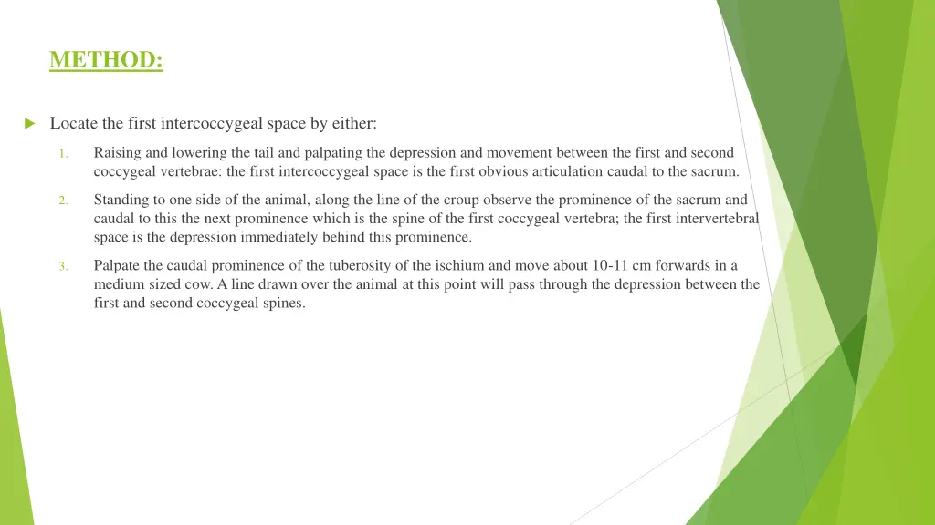
METHOD: Locate the first intercoccygeal space by either: Raising and lowering the tail and palpating the depression and movement between the first and second coccygeal vertebrae: the first intercoccygeal space is the first obvious articulation caudal to the sacrum. 1. Standing to one side of the animal, along the line of the croup observe the prominence of the sacrum and caudal to this the next prominence which is the spine of the first coccygeal vertebra; the first intervertebral space is the depression immediately behind this prominence. 2. Palpate the caudal prominence of the tuberosity of the ischium and move about 10-11 cm forwards in a medium sized cow. A line drawn over the animal at this point will pass through the depression between the first and second coccygeal spines. 3.
To administer the local anesthesia either of two methods can be used: Hanging drop or Lack of resistance technique. First, disinfect the skin over the first intercoccygeal space using 70% Isopropyl alcohol. Inject a small amount of 2% Lidocaine; local anaesthetic to desensitise the skin over the injection site and minimise reaction during insertion of the needle. https://www.youtube.com/watch?v=rbe9B5tFBF0
An 18-Gauge, 1 1/2 needle is used to penetrate the intervertebral space . The needle is usually directed slightly at 45 in a cranial direction and advanced slowly ventrally and cranially until the needle touches the floor of the spinal canal. A popping sensation may be felt as the needle penetrates the dura. Correct placement of the needle can be checked by the Hanging drop technique which can be performed by placing few drops of sterile water or lidocaine into the needle hub during insertion. When the needle enters the correct space, the drop of Lidocaine is observed to be aspirated under the effect of the negative pressure in the epidural space. This is called the Lack of resistance technique. Furthermore before injection of the drug, negative pressure is applied by the syringe to ensure blood or spinal fluid is not aspirated. In which case, the needle must be withdrawn and adjusted slightly and negative pressure is applied again.