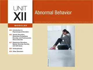
Commonly Seen Skin Disorders in School-Aged Children
This presentation covers infectious and non-infectious skin disorders in school-aged children, including anatomy and physiology of the skin, assessment strategies, and management approaches. It emphasizes the importance of history collection, discusses common skin disorders affecting children, and provides insights on Acanthosis Nigricans.

Download Presentation

Please find below an Image/Link to download the presentation.
The content on the website is provided AS IS for your information and personal use only. It may not be sold, licensed, or shared on other websites without obtaining consent from the author.If you encounter any issues during the download, it is possible that the publisher has removed the file from their server.
You are allowed to download the files provided on this website for personal or commercial use, subject to the condition that they are used lawfully. All files are the property of their respective owners.
The content on the website is provided AS IS for your information and personal use only. It may not be sold, licensed, or shared on other websites without obtaining consent from the author.
E N D
Presentation Transcript
Commonly Seen Infectious and Non- Infectious Skin Disorders in the School- Aged Child Melinda Rodriguez DNP, APRN, FNP-BC Nursing Education Doctor s Hospital at Renaissance Health Systems
Disclosures Nothing to disclose
Objectives Discuss a brief overview of the anatomy and physiology of the skin Discuss the importance of history collection Discuss infectious skin disorders affecting the school-aged child Discuss the non-infectious skin disorders affecting the school-aged child Discuss assessment and management of Acanthosis Nigricans (AN)
Brief Overview of Integumentary System Provides an elastic, rugged, self-regenerating cover for the body Largest organ of the body Includes: hair and nails Maintains and keeps body structures in place
Anatomy and Physiology Comprised of several layers Protects against microbial and foreign substance invasion Regulates body temperature Provides sensory perception via nerve endings Produces vitamin D from precursors in skin Contributes to blood pressure regulation
Functions of the Skin Complex organs made up of may cell types Largest organ of the body Provides barrier between external and internal environments Provides protection against organisms Skin receptors relay: touch, pressure, temperature and pain to CNS Also provide ability for localization and discrimination McCance & Huether, (2014).
Overview of the Skin Assessment Problems may arise from many mechanisms and inflammatory processes Some causes may be environmental, traumatic and secondary to exposures Evaluation of skin disorders require a in-depth focus history and PE Assess for infectious symptoms: fever, itching Look at the presentation of lesion, configuration and distribution Seidel, Ball, Dains et al., (2015).
External Clues to Internal Problems Persistent pruritus may indicate chronic renal failure, liver disease, diabetes Supernumerary nipples located along mammary ridge, may be associated with renal problems Facial port wine stain may be associated with ocular defects, malformation of meninges
Age-Appropriate History Gather data specific to current skin problems Family, PMH of similar problems Skin care routines Recent changes in skin, hair or nail care Sun-exposure habits; use of sunscreen Medication history Onset, date of occurrence History of recent travel Rx medications; OTC medications, lotions used
History of Present Illness Note recent or past changes in the skin: pruritus, dryness, sores, rashes, lumps Symptoms: pain, exudate, bleeding, color changes Recent drug exposure; chemicals; Generalized symptoms: fever, travel hx Use of topical or oral medications Seidel et al., (2015).
History (contd) Eating habits; allergies to foods Communicable disease exposure Allergic disorders; asthma Exposure to pets; animals Skin injury; outdoor exposures Nail biting Thinning of hair Seidel et al., (2015).
Mechanisms of Self-Defense Bacteria-Derived Chemicals: skin, mucous membranes and GI tract, urethra and vagina have protective microorganisms Common bacteria on the skin: staph and strept C-difficile in the GI tract Lactobacillus protection of the vaginal tract
Inspection of the Skin Performed by inspection and palpation Inspection: lighting is essential Observe for symmetry Adequate exposure of the skin Inspect skin thickness Assess for color variances Assess for nevi; abnormally shaped; variegated colors Seidel et al., (2015).
Palpation of the Skin Palpate for the following: Moisture Temperature Texture Turgor Mobility Image result for skin palpation Visualdex.com Seidel et al., (2015).
Blood supply/nerve innervation Blood supply to skin limited Include papillary capillaries Dermis facilitates the regulation of body temperature Evaporation of sweat cools body Regulates vasoconstriction McCance & Huether, (2010).
Morphological Criteria Includes: Location of lesion Distribution Determine whether primary or secondary Shape of lesion Margins/borders/irregularities Pigmentation/color/variations Palpate texture/consistency Wear gloves if open lesions present Seidel et al., (2015).
Morphological Characteristics of Lesions Linear (in a line) Stellate (star shaped) Reticulate (netlike; lacy) Mobilliform (maculopapular; confluent) Irregular borders Border raised above Advancing; spreading beyond borders (cellulitis) Seidel et al., (2015).
Pigmentation Flesh colored Erythematous/pink Salmon colored (psoriasis) Black Purple Yellow/waxy Pearly
Primary Skin Lesions Macule: flat, circumscribed area; changes to color of skin; less than 1cm in diameter (freckle) Papule: elevated firm circumscribed area less than 1cm (wart) Patch: a flat non-palpable irregular shaped macule; more than 1cm (vitiligo) Plaque: elevated, firm, rough with flat top surface; greater than 1cm in diameter ( psoriasis) Vesicle: elevated, circumscribed superficial; does not extend to dermis, filled with serous fluid less than 1cm McCance & Huether, (2014).
Primary Skin Lesions Macule/Papule
Secondary Lesions (contd) Scale: heaped up keratinized flaky skin; thick or thin, dry variation in size (seborrheic dermatitis) Lichenification: rough, thickened epidermis secondary to persistent rubbing, itching of skin; flexor surfaces of skin (chronic dermatitis) Scar: thin to thick fibrous tissue; replaces normal skin following injury (healed wound) Keloid: irregular-shaped, elevated progressively enlarging, goes beyond boundaries of the wound; excessive collagen formation McCance & Huether, (2014).
Secondary Lesions Image result for keloid Image result for scar Scar Keloid medicinenet.com
Vascular Skin Lesions Spider angioma; red central body with spider-like legs; blanches with pressure Purpura; is red purple in color; non-blanchable; greater than 0.5cm in diameter Petechiae; red-purple in color, non-blanchable; less than 0.5cm in diameter Telangiectasia; fine, irregular red lines Venous star; bluish spider; irregular shape does not blanch with pressure
Vascular Lesions Telangiectasias
Pigment Disorders of the Skin Skin reflects emotional states Warmth and other responses are given/received Pigmentary skin disorder: vitiligo affects people of all races, sudden appearances of white patches; vary in size, hereditary and genetic cause Albinism: genetic disorder absence of pigment in skin, hair, eyes; found in all races Melasma: darkened macules on face; OC use; exacerbated by sun exposure McCance & Huether, (2010).
Assessment of the Adolescent Increased oiliness or perspiration may be evident Increased axillary perspiration related to maturity of the apocrine glands Hair on extremities becomes coarser and darker Pubic hair develops; secondary sex characteristics
Infectious and Non-Infectious Conditions of the Skin Management and Treatment
Common Skin Disorders Seen in the Schools Impetigo Varicella Scabies/Pediculosis Herpes simplex Contact dermatitis/eczema Molluscum Contagiosum Hand, Foot and Mouth Disease Fifth s Disease (erythema infectiosum) Rubeola /Measles Stept Infection (Scarletina)
Infectious vs. Non-Infectious History of present illness is very important Events that preceded the skin condition Need to rule out trauma Medication history Previous outbreak Fever and any other systemic symptoms Allergies
Eczema Characterized by : acute inflammation, erythema, edema and vesiculation Itching is often severe Multiple causes; allergic contact Common culprits: personal care products, fragrances, detergents Often sudden in onset Habif, (2011).
Prognosis/Management Avoid provoking factors; eruption improves in 7-10 days Excoriation secondary to itching/scratching could develop bacterial infection Topical steroids (used sparingly and as directed) Oral antihistamines (Benadryl) Treatment often based on elimination of causing factor Habif, (2011).
Allergic Contact Dermatitis Common T-cell mediated or delayed hypersensitivity Allergens: chemicals, foreign proteins, poison ivy Erythema, swelling with itching Vesicular lesions are where contact is made Removal is necessary to help with tissue repair Systemic steroids are one form of treatment Atopic dermatitis: more common in infancy and childhood, usually associated with asthma, allergic rhinitis McCance & Huether, (2014).
Allergic Contact Dermatitis Delayed type hypersensitivity reaction Caused by skin contact with an allergen Results in eczematous dermatitis Common causes include: Metals (nickel) Rubber Shoes Preservatives in lotions, creams, cosmetics Habif, (2011).
Allergic Contact Dermatitis Image result for allergic contact dermatitis is due to Pathologyoutlines.com Mayoclinic.org
Management/ Treatment Avoidance of the allergenic substance Identification of allergen (patch testing) Topical treatment (topical corticosteroids) Choice of topical corticosteroids depends on body site affected (use sparingly on pediatric population) 3-week tapering course of oral corticosteroids Education of patient/caregiver Habif, (2011).
Pediatric Considerations Allergies can develop after years of exposure to products/medications Consider patch testing Re-assessment of recent exposures Assess the integrity of the skin Be alert for S/S of infection Habif, (2011).
Bacterial Infections of the Skin Can result from primary skin lesions Any break in the integrity of the skin May result in erythema, edema, pain, pus May result in systemic symptoms such as: Fever Malaise Myalgias Nausea and vomiting
Impetigo Highly contagious superficial skin infection Caused by strept or staph 80% of cases caused by staph aureus Occurs after minor skin injury, insect bite Bacteria may colonize in the nasal passages Warm climates and poor hygiene contribute to it Lesions may be localized or wide spread; common on face Habif, (2011).
Skin Findings Vesicles/pustules present Red a moist base Erythematous Lesions often coalesce Develop an adherent crust honey-yellow to white- brown in color Thin-roofed bullae may develop Habif, (2011).
Impetigo Image result for impetigo pictures OSC Microbio 21 02 impetigo.jpg Medicinenet.com
Pediatric Considerations Most common bacterial infection in children Rarely post-streptococcal glomerulonephritis may follow infection Antibacterial soaps are recommended to be used twice daily for chronic cases Bacterial culture may be indicated for chronic cases Habif, (2011).
Treatment/Management Disease is self-limiting; could spread Localized infections: Mupirocin 2% topical Oral antibiotics: doxycycline, clarithromycin, cephalexin (Keflex) x 10-14 days of treatment Recurrent impetigo may require topical Mupirocin in the nares Good handwashing Habif, (2011).
Viral Infections Verucca: warts, common benign papillomas; caused by HPV; transmitted by direct contact Herpes simplex: (HSV) infection of skin and mucous membranes; two types HSV 1 and HSV 2; symptoms begin with burning or tingling; umbilicated vesicles and erythema Herpes Zoster: shingles; acute localized vesicular eruption distributed along dermatomal segment; prevention via Zostavax vaccine McCance & Huether, (2014).
Verruca Vulgaris Also known as warts Benign epidermal proliferations Caused by human papilloma virus (HPV) Over 150 different types of HPV Transmission is by simple contact; often on non-intact skin Local spread is caused by autoinoculation Peak incidence ages 12-16 yrs Habif, (2011).
Skin Findings Flesh-colored papules evolve into dome shaped, gray to brown, hyperkeratotic , rough papules Common sites: Hands Skin Periungual Knees, plantar surfaces Habif, (2011).
Management/Treatment Course is highly variable Spontaneous resolution with time 2/3 of warts in children regress within 2 years Multiple treatments are available OTC topical salicylic acid preparations Duration of treatment is usually 8-12 weeks Cryotherapy Imiquimod 5% cream (Aldara) Habif, (2011).
Herpes Simplex Double-stranded DNA virus; two virus types (types 1 & 2) Type I associated with vesicular, ulcerative oral infections Type II associated with genital infections Primary infection can be asymptomatic Spread by respiratory droplets, direct contact with active lesion Contact with virus containing fluid: saliva, cervical secretions in people with no active disease Symptoms occur 3-7 days after contact Habif, (2011).
Herpes Simplex I & II (HSV-1 and HSV2) Image result for herpes simplex virus Image result for herpes simplex virus Image result for herpes genital Clinicaladvisor.org
Primary Infection Tenderness, pain, mild paresthesias or burning before onset of lesion Grouped vesicles on erythematous base appear; subsequently erode Lesions on the mucus membrane accumulate exudate; on skin may form a crust Lesions last 2-6 weeks and heal without scarring Habif, (2011).
Recurrent Infection Recurrence rate is same as primary infection Local skin trauma, systemic changes (fatigue, fever) reactivate the virus Travels down the peripheral nerve to site of initial infection Prodromal symptoms may last 2-24 hours Many can experience a decrease in outbreaks with time Habif, (2011).


")


")


")