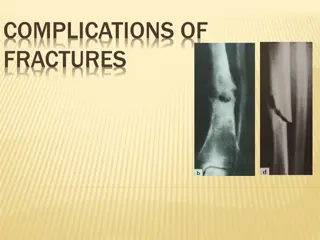
Complications of Rhinosinusitis: Symptoms, Spread, and Classification
Learn about the complications of rhinosinusitis, including symptoms, routes of spread, and classification. Understand the potential orbital and intracranial complications that can arise from this condition. Find out about cavernous sinus thrombosis, treatment strategies, and common clinical features. Explore the various types of infections that can lead to osteomyelitis in the maxilla or frontal bone.

Download Presentation

Please find below an Image/Link to download the presentation.
The content on the website is provided AS IS for your information and personal use only. It may not be sold, licensed, or shared on other websites without obtaining consent from the author. If you encounter any issues during the download, it is possible that the publisher has removed the file from their server.
You are allowed to download the files provided on this website for personal or commercial use, subject to the condition that they are used lawfully. All files are the property of their respective owners.
The content on the website is provided AS IS for your information and personal use only. It may not be sold, licensed, or shared on other websites without obtaining consent from the author.
E N D
Presentation Transcript
Complications of Rhinosinusitis BY Mahmood A. Hamed OTORHINOLARYNGOLOGY Department
Definition: As long as infection is confined only to the sinus mucosa, it is called sinusitis. Complications are said to arise when infection spreads into or beyond the bony wall of the sinus. Alarming Signs:- 1- Progression of symptoms and deterioration of general condition . 2- Appearance of new symptoms and signs (Proptosis, external swelling, deterioration of vision, positive meningeal signs, neurologic deficit, Disturbed conscious level, ..etc.
Routes of spread: 1- Direct 2- Venous 3- Lymphatic 4- Perineural
Classfication : A- Cranial Osteomyelitis- frontal bone and maxilla B- Orbital Preseptal inflammatory oedema of lids Subperiosteal abscess Orbital cellulitis Orbital abscess Superior orbital fissure syndrome Orbital apex syndrome Meningitis Extradural abscess Subdural abscess Brain abscess Cavernous sinus thrombosis C- Intacranial D- Descending infections
Cavernous sinus thrombosis : Cavernous sinus thrombosis : Aetiology Aetiology: : Infection of paranasal sinuses and orbital complications from these sinus infections can cause thrombophlebitis of the cavernous sinus. Clinical features: Clinical features: Abrupt onset with chills and rigors. Swollen eyelids with chemosis and proptosis of eyeball. CN III, IV and VI get involved individually and sequentially causing total ophthalmoplegia. Pupil becomes dilated and fixed. Congestion of optic disc with diminution of vision. Sensation in the distribution of V is diminished. Treatment: Treatment: i.v. antibiotics and attention to the focus of infection, drainage of infected ethmoid or sphenoid sinus. 1
Posterior group of paranasal sinuses can give rise to: 1- Superior orbital fissure syndrome. 2- Orbital apex syndrome.
. OSTEOMYELITIS Osteomyelitis is infection of bone marrow and should be differentiated from osteitis which is infection of compact bone. It involves either maxilla or frontal bone. 1. Osteomyelitis of maxilla More often seen in infants and children because of presence of spongy bone in the anterior wall of the maxilla. Clinical features: Erythema, swelling of cheek, lower lid oedema, purulent nasal discharge and fever. Subperiosteal abscess followed by fistulae may form in infraorbital region,alveolus, or in zygoma. Sequestration of bone may occur. Treatment: Large doses of antibiotics, drainage of any abscess and sequestra removal.
2. Osteomyelitis of frontal bone More often seen in adults as frontal sinus is not developed in infants and children. It may result from acute infection of frontal sinus either directly or through the venous spread. Pus may form externally under the periosteum as soft doughy swelling ( (Pott s Pott s puffy puffy tumour tumour), ), or internally as an extradural abscess. Treatment: Large doses of antibiotics, drainage of abscess and trephining of frontal sinus through its floor. Sometimes, it requires removal of sequestra and necrotic bone by raising a scalp flap through a coronal incision.
INTRACRANIAL COMPICATIONS Frontal, ethmoid and sphenoid sinuses are closely related to anterior cranial fossa and infection from these can cause following complications: 1) Meningitis and encephalitis 2) Extradural abscess 3) Subdural abscess 4) Brain abscess 5) Cavernous sinus thrombosis
THANK YOU